Download
1 / 128
1.32k likes | 1.56k Views
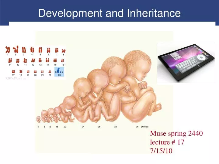
Development and Inheritance. Muse spring 2440 lecture # 17 7/15/10. Development. Differentiation Creation of different types of cells required in development Occurs through selective changes in genetic activity As development proceeds, some genes are turned off, others are turned on

E N D
Development and Inheritance Muse spring 2440 lecture # 17 7/15/10
Development • Differentiation • Creation of different types of cells required in development • Occurs through selective changes in genetic activity • As development proceeds, some genes are turned off, others are turned on • Fertilization • Also called conception • When development begins
Development • Embryological Development • Occurs during first 2 months after fertilization • Study of these events is called embryology • Fetal Development • Begins at start of ninth week • Continues until birth
Development • Prenatal Development • Embryological and fetal development stages • Postnatal Development • Commences at birth • Continues to maturity when aging process begins
Fertilization • Fertilization • Fusion of two haploidgametes, each containing 23 chromosomes • Produces zygote containing 46 chromosomes Fertilization and the Preparation for Cleavage
Fertilization • Gamete • Provides • Cellular organelles (female) • Inclusions • Nourishment • Genetic programming necessary to support development of embryo for a week
Fertilization • Fertilization • Occurs in uterine tube within a day after ovulation • Secondary oocyte travels a few centimeters • Spermatozoa must cover distance between vagina and ampulla (30 + cm)
Fertilization • Hyaluronidase • Enzyme breaks down bonds between adjacent follicle cells • Allows spermatozoon to reach oocyte • Acrosin • Is a proteolytic enzyme • Is required to reach oocyte
Fertilization • Acrosomal Caps • Release hyaluronidase and acrosin • Penetrate corona radiata, zona pellucida, toward oocyte surface • Oocyte Activation • Contact and fusion of cell membranes of sperm and oocyte • Follows fertilization • Oocyte completes meiosis II, becomes mature ovum
Fertilization • Polyspermy - would be bad • Fertilization by more than one sperm • Prevented by cortical reaction • Cortical Reaction- initiated upon sperm penetration • Releases enzymes that • Inactivate sperm receptors • Harden zona pellucida • Lift fertilization envelope (vitelline layer)
Fertilization • Female Pronucleus • Nuclear material remaining in ovum after oocyte activation • Male Pronucleus • Swollen nucleus of spermatozoon • Migrates to center of cell
Fertilization • Amphimixis • Fusion of female pronucleus and male pronucleus • Moment of conception • Cell becomes a zygote with 46 chromosomes • Fertilization is complete
Fertilization • Cleavage • Series of cell divisions • Produces daughter cells • Differentiation • Involves changes in genetic activity of some cells but not others
Fertilization Figure 29–1a Fertilization: An Oocyte and Numerous Sperm at Time of Fertilization.
Fertilization Figure 29–1b Fertilization and the Preparations for Cleavage.
Fertilization Figure 29–1b Fertilization and the Preparations for Cleavage.
Fertilization Figure 29–1b Fertilization and the Preparations for Cleavage.
Fertilization Figure 29–1b Fertilization and the Preparations for Cleavage.
Gestation • Induction • Cells release chemical substances that affect differentiation of other embryonic cells • Can control highly complex processes • Gestation • Time spent in prenatal development • Consists of three integrated trimesters, each 3 months long
Gestation • First Trimester • Period of embryological and early fetal development • Rudiments of all major organ systems appear • Second Trimester • Development of organs and organ systems • Body shape and proportions change • By end, fetus looks distinctively human • Third Trimester • Rapid fetal growth and deposition of adipose tissue • Most major organ systems are fully functional
The First Trimester • Cleavage • Sequence of cell divisions begins immediately after fertilization • Zygote becomes a pre-embryo, which develops into multicellular blastocyst • Ends when blastocyst contacts uterine wall
The First Trimester • Implantation • Begins with attachment of blastocyst to endometrium of uterus • Sets stage for formation of vital embryonic structures • Placentation • Occurs as blood vessels form around periphery of blastocyst and placenta develops
The First Trimester • Placenta • Complex organ permits exchange between maternal and embryonic circulatory systems • Supports fetus in second and third trimesters • Stops functioning and is ejected from uterus after birth • Embryogenesis • Formation of viable embryo • Establishes foundations for all major organ systems
The First Trimester • Most dangerous period in prenatal life • 40% of conceptions produce embryos that survive past first trimester
The First Trimester • Blastomeres • Identical cells produced by cleavage divisions • Morula • Stage after 3 days of cleavage • Pre-embryo is solid ball of cells resembling mulberry • Reaches uterus on day 4
The First Trimester Figure 29–2 Cleavage and Blastocyst Formation.
The First Trimester • Blastocyst • Formed by blastomeres • Hollow ball with an inner cavity • Known as blastocoele
The First Trimester • Trophoblast • Outer layer of cells separate outside world from blastocoele • Cells responsible for providing nutrients to developing embryo
The First Trimester • Inner Cell Mass • Clustered at end of blastocyst • Exposed to blastocoele • Insulated from contact with outside environment by trophoblast • Will later form embryo
The First Trimester Figure 29–2 Cleavage and Blastocyst Formation.
The First Trimester • Implantation • Occurs 7 days after fertilization • Blastocyst adheres to uterine lining • Trophoblast cells divide rapidly, creating several layers Stage of Implantation
The First Trimester • Cellular Trophoblast • Cells closest to interior of blastocyst • Syncytial Trophoblast • Outer layer • Erodes path through uterine epithelium by secreting hyaluronidase
The First Trimester Figure 29–3 Stages in Implantation.
The First Trimester • Ectopic Pregnancy • Implantation occurs outside of uterus • Does not produce viable embryo • Can be life threatening • Lacunae • Trophoblastic channels carrying maternal blood
The First Trimester • Villi • Extend away from trophoblast into endometrium • Increase in size and complexity until day 21 • Amniotic Cavity • A fluid-filled chamber • Inner cell mass is organized into an oval sheet two layers thick • Superficial layer faces amniotic cavity • Deeper layer is exposed to fluid contents of blastocoele
The First Trimester • Gastrulation • Formation of third layer of cells • Cells in specific areas of surface move toward central line • Known as primitive streak
Gastrulation Week 3 - 15 days in
The First Trimester • Primitive Streak • Migrating cells leave surface and move between two layers • Creates three distinct embryonic layers, or germ layers • Ectoderm: consists of the superficial cells that did not migrate into interior of inner cell mass • Endoderm: consists of cells that face blastocoele • Mesoderm: consists of poorly organized layer of migrating cells between ectoderm and endoderm
The First Trimester Ectoderm makes me nervous
The First Trimester Mesoderm is myo favorite
The First Trimester Endoderm gives me endogestion
The First Trimester • Embryonic Disc • Oval, three-layered sheet • Produced by gastrulation • Will form body of embryo • Rest of blastocyst will be involved in forming extraembryonic membranes
The First Trimester Figure 29–4 The Inner Cell Mass and Gastrulation.
The First Trimester • Formation of the Extraembryonic Membranes • Support embryological and fetal development • Yolk sac • Amnion • Allantois • Chorion
The First Trimester • Yolk Sac • Begins as layer of cells spread out around outer edges of blastocoele to form complete pouch • Important site of blood cell formation • Amnion • Combination of mesoderm and ectoderm • Ectodermal layer enlarges and cells spread over inner surface of amniotic cavity • Mesodermal cells create outer layer • Continues to enlarge through development
The First Trimester • Amniotic Fluid • Contained in amniotic cavity • Surrounds and cushions developing embryo or fetus • Allantois • Sac of endoderm and mesoderm • Base later gives rise to urinary bladder
The First Trimester • Chorion • Combination of mesoderm and trophoblast • Blood vessels develop within mesoderm • Rapid-transit system for nutrients that links embryo with trophoblast • First step in creation of functional placenta
The First Trimester • Chorionic Villi • In contact with maternal tissues • Create intricate network within endometrium carrying maternal blood • Body Stalk • Connection between embryo and chorion • Contains distal portions of allantois and blood vessels that carry blood to and from placenta
The First Trimester • Yolk Stalk • Narrow connection between endoderm of embryo and yolk sac • Decidua Capsularis • Thin portion of endometrium • No longer participates in nutrient exchange and chorionic villi in region disappear
The First Trimester Figure 29–5 Extraembryonic Membranes and Placenta Formation.