Download
1 / 61
620 likes | 661 Views
DISEASES OF THE BLOOD VESSELS. ATHEROSCLEROSIS. Dr Eman MS Muhammad. DISEASES OF THE BLOOD VESSELS. Diseases of arteries Degenerative Inflammatory Obliterative Aneurysms Diseases of veins. Diseases of Arteries. Arteriosclerosis: Atherosclerosis

E N D
DISEASES OF THE BLOOD VESSELS ATHEROSCLEROSIS Dr Eman MS Muhammad
DISEASES OF THE BLOOD VESSELS • Diseases of arteries • Degenerative • Inflammatory • Obliterative • Aneurysms • Diseases of veins
Diseases of Arteries • Arteriosclerosis: • Atherosclerosis • Monkeberg’s medial calcific sclerosis • Arteriolosclerosis • Inflammations (arteritis- vasculitis or vasculitides), polyarteritisnodosa (PNA), Buerger’s disease, giant cell arteritis, Wegener’s granulomatosis, ……etc • Aneurysms.
Terminology • The following terms are similar, yet distinct, in both spelling and meaning, and can be easily confused: arteriosclerosis, arteriolosclerosis, and atherosclerosis. • Arteriosclerosisis a general term describing any hardening (and loss of elasticity) of medium or large arteries. • Arteriolosclerosisis any hardening (and loss of elasticity) of arterioles (small arteries).
Atherosclerosisis a hardening of an artery specifically due to an atheromatous plaque. • The short term atheroma has historical priority and is less easily confused with the quite separate process of arteriolosclerosis (hypertension). • Atherogenic is used for substances or processes that cause atherosclerosis.
Atherosclerosis (Atheroma) • Definition: Atherosclerosis (AS)is acommon degenerative disease in which patchy deposits of fatty material develop in the walls of medium-sized and large arteries, followed by fibrosis, leading to reduced blood flow. • It is a slow complex process in which fatty substances, cholesterol, cellular waste products, and calcium build up in the inner lining of an artery.
This buildup is called plaque. • AS is derived from the Greek words athero (meaning gruel or paste) and sclerosis (meaning hardness). • Recently, atherosclerosis is defined as a chronic multifocal immuno-inflammatoryfibroproliferative disease affecting large and middle-sized arteries, mainly induced by lipids accumulation. • It is always present in some degree in middle-aged and old people, causing narrowing of the lumen of the arteries.
In most developed industrialized Western countries atheroma is the main cause of disability and death from heart diseases, cerebral infarction and of ischemia of the lower limbs and gut.
Macroscopic picture: • The earliest deposits of lipids in the intima of the aorta and large arteries occur in childhood and adolescence. They are known as “fatty streaks”. • They appear as yellow slightly raised areas on the luminal surface, which enlarge and coalesce to form irregular yellow streaks. • They consist of accumulation of lipid droplets beneath the endothelium, both free and as aggregates within the macrophages.
Other lesions which may precede atheroma include: • Foci of intimal edema, and ↑ in the ground substance and smooth muscle cells;“intimal cushions” occur at the branching points of the arteries. • Small disc like slightly raised patches “plaques”of intimal thickening with smooth glistening surface. • The patches enlarge and thicken by further deposition of lipids deep in the intima and by fibrosis more superficially.
When viewed from the intimal surface these raised plaques appear yellow or white depending on the amount of fibrous tissue overlying the lipid deposits. • The patches vary in thickness, reflecting their development and slow growth throughout life.
Microscopic picture: • The earliest changes are due to proliferation of smooth muscle cells in the intima and accumulation of lipids in “foam cells”. • Some foam cells are macrophages; derived from monocytes which adhere to and then penetrate the endothelium. • Others are medial smooth muscles which proliferate and migrate into the intima. • Both absorb lipids and their cytoplasm becomes swollen with lipid globules.
Extra-cellular lipids accumulate in the intima near to the media in relation to elastic fibers and the internal elastic lamina. • As the patches develop, thin strands of CT appear sub-endothelially and between foam cells and form the fibrous part of the lesion “fibrous cap”. • Necrosis develop in the deeper part of the lesion with accumulation of extracellular lipids, cholesterol crystals and tissue debris. • Infiltration by PNL and other inflammatory cells is common.
The internal elastic lamina is disrupted. • Necrosis and fibrosis erode media, and as the plaques thicken, the underlying media becomes thin and atrophic. • Small blood vessels grow into the atheromatous plaque from the vasavasorum. • They may hemorrhage contributing altered blood constituents to the plaque contents. • Some plaques may crack and rupture causing hemorrhage into the plaque leading to the formation of thrombus.
Clinical effects: • They vary depending on the size of the artery involved: • Uncomplicated atheroma of large arteries usually has no clinical effectsbecause it doesn’t reduce the lumen seriously. • In advanced cases an aneurysm may form. • Mural thrombus seldom causes complete occlusion of the aorta except at or near its bifurcation, with resulting coldness and weakness of the legs, muscle wasting, and sexual impotence.
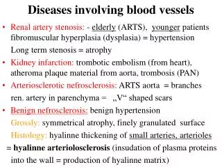
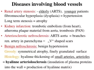
Fragments of thrombi and athermanous debris from ulcerated plaques may form emboli which lodge in arteries of the legs, gut, kidneys and spleen. • The most important effect of atheroma is due to involvement of smaller arteries. The lumen may be progressively narrowed by an atheromatous plaque causing chronic ischemia or suddenly occluded by thrombosis which often causes infarction.
Thrombosis is often precipitated by a crack in the fibrous cap. The most dangerous effect is the coronary artery thrombosis. Atheroma is the chief cause of ischemic heart disease, the largest single cause of death in developed countries. • Atheroma also is a very common cause of cerebrovascular diseases, due to atheroma of the carotid, vertebral and cerebral arteries. • Aneurysm may develop in the circle of Willis.
Peripheral vascular diseaseis also common especially in diabetics, and cigarette smokers. Intermittent claudication painproduced in the leg due to muscle ischemia induced by exercise and relieved by rest. • If ischemia is severe gangrene may develops which starts in the toes and spreads proximally.
Clinical complication of atheroma: • Ischemic heart diseases: • Sudden death • Angina pectoris • Myocardial infarction • Cardiac arrhythmias • Cardiac failure
Cerebral ischemia • Transient cerebral ischemic attack • Dementia • Cerebral infarction • Mesenteric ischemia • Abdominal claudication • Malabsorption syndrome • Bowel infarction
Peripheral vascular disease • Intermittent claudication pain • Gangrene • Renovascularhypertention • Renal artery stenosis • Aneurysms • Aorta • Iliac and popliteal arteries • Cerebral arteries specially “Circle of Willis”
Atheroma& Lipid Metabolism • Chylomicrons • Transport lipid from intestine to liver • VLDL • Carry cholesterol and TG from liver • TG removed leaving LDL • LDL • Rich in cholesterol • Carry cholesterol to non-liver cells • HDL • Carry cholesterol from periphery back to liver
Lipids circulate in plasma in combination with proteins in the form of lipoproteins. • Type of lipoprotein Type of lipid • Chylomicrons………. Triglycerides (90%) • HDL………….. Cholesterol (<20%) • LDL…………... Cholesterol (75%) • VLDL…………. Cholesterol (25%)
Familial Hyperlipidemia • Genetically determined abnormalities of lipoproteins. • Lead to early development of atheroma. • Associated physical signs: • xanthelasma a sharply demarcated yellowish deposit of fat underneath the skin, around the eyelids which may be referred to as a xanthoma when becoming larger and nodular.
Age: • AS is not manifested in children. The incidence and severity ↑as the age advances. • Clinically manifests in late middle or old age. • Sex: • Higher incidence in males. Estrogen ↓ the incidence in females up to the age of menopause. • Before 45ys ♂:♀ 5:1 • After 45ys ♂:♀ 1:1
Stress: • Predisposes to AS as evident by: • Hypertension: ↑ the incidence and complications of AS. • AS is more common around the mouths of the intercostal and lumbar arteries because of the stress caused by the turbulent blood flow. • Diet: • Excess intake of animal fats favors the development ofAS.
Plasma lipids: • High level of LDL is an important factor in the development of AS. • Hypercholesterolemia: • It is a major risk factor as evidenced by: • AS plaque is rich in cholesterol. • Genetically induced hypercholesterolemia → premature AS.
Increased mortality of IHD in persons having high plasma cholesterol level. • Treatment with diet or drugs that lowering cholesterol →↓ in death rate of IHD in hypercholesterolemic patient. • High dietary intake of cholesterol and saturated fatty acids (FA) → ↑plasma cholesterol level. • N.B. Omega-3 FA (in fish & fish oils) → ↓ plasma level of LDL and ↑HDL level.
Artificial trans fats (or trans fattyacids) are created in an industrial process that adds hydrogen to liquid vegetable oils to make them more solid. • The primary dietary source for trans fatsin processed food is “partially hydrogenated oils.“ They have a very important role in AS development.
Hypertension: • Increased blood pressure → acceleration of AS &↑incidence of IHD and cerebrovascular diseases. • Anti-hypertensive →↓ incidence of strokes and IHD. • Diabetes mellitus: • Both type I & II diabetes are associated with 2 folds ↑ risk of AS, myocardial infarction, cerebral infarction and gangrene of lower limbs. • Heredity: • Familial tendency is well known possibly due to: • variations in apolipoprotein metabolism. • variations in apolipoprotein receptors.
Cigarette smoking: • Death rate from IHD is 70-200% higher in smokers than in non-smokers. • Quitting cigarette smoking →↓ risk of death from IHD. • Mode of action is uncertain : • Disruption of coagulation system • reduced PGI2 • increased platelet aggregation
Others (Soft risk factors): • Insufficient regular physical activity • Competitive stressful lifestyle with type A person • Obesity • Oral contraceptives • Hyperuricemia • High carbohydrate intake
Pathogenesis: • Old theories: • Insudation theory: • Excess intake of animal fats causes hypercholesterolemia and increased plasma beta lipoproteins. • Plasma passes constantly from the vascular lumen through the vessel wall.
Repeated endothelial injury↑ endothelial permeability to plasma lipoproteins →large beta lipoproteinmolecules are trapped in the subintimal connective tissue. • The protein part is filtered, while the insoluble lipid part is retained and induces fibrosis around.
Thrombogenic/encrustation theory: • It supposed that small thrombi collected over foci of endothelial injury (initial event) and organization of such thrombi resulted in plaque formation. • Llipid is derived from breakdown of platelets and leucocytes rather than serum lipoprotein. • Endothelial injury may be due to: aging process, hypertension, viruses, stress, cigarette smoking or immune complex deposition.
Recent theory: • Reaction to injury theory: • This theory suppose that AS is a response to endothelial injury. • It was the most accepted theory till recently. • Original theory: • Initial event in AS is endothelial injury followed by smooth muscle cell proliferation. • This theory suggests that the early lesions mainly consist of smooth muscle cells proliferation.
Modified theory: • Describes lipoprotein entry into the intima as the initial event followed by lipid accumulation in the macrophages (foam cells) which according to the modified theory are the dominant cells in early lesions.
Response to injury hypothesis suppose that: • Injury may be: • Gross e.g. physical, chemical traumas. • Subtle e.g. hypertension, diabetes, hyperlipidemia, cigarette smoking. • Injury →↑ permeability to plasma constituent → platelet and monocyte adherence to endothelium. • Platelets and monocytes release factors (PDGF) → smooth muscle migration from the media to the intima followed by proliferation.
Smooth muscle synthesize ECM. • Smooth muscle and macrophages accumulate lipids → formation of foamy cells. • Macrophages release enzymes, cytokines (IL-1 &TNF) and oxidants → oxidation of LDL → further injury.
Response to injury hypothesis in the development of atherosclerosis