Download

1 / 17
170 likes | 271 Views
Vogt-Koyanagi Harada Disease. Fernando Oréfice Juliana Lambert Oréfice Centro Brasileiro de Ciências Visuais- Brazil. Ocular History. 31 year woman Sudden blurred vision from 2 days prior. First Presentation. VA: OD 20/200, OS 20/400 Mild AC cell 1+/4+ Fundus: Hyperaemia of disc

E N D
Vogt-Koyanagi Harada Disease Fernando Oréfice Juliana Lambert Oréfice Centro Brasileiro de Ciências Visuais- Brazil
Ocular History • 31 yearwoman • Sudden blurredvisionfrom 2 daysprior
First Presentation • VA: OD 20/200, OS 20/400 • Mild AC cell 1+/4+ • Fundus: • Hyperaemiaofdisc • Exudativeretinaldetachment
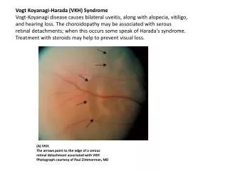
Serous detachment • Hyperemia disk
Spectral OCT Serous detachment Serous detachment
Spectral OCT • Posterior retinal irregular profile. • Macular serous detachment
Fundusfluoresceinangiography OD: disc hyperfluorescence OS pinpoint
Diagnosis • Vogt-Koyangi Harada Disease • Involvement of the optic nerve • very significant • justifies a more aggressive treatment.
Treatment • Intravenousmethyprednisolone • 250mg IV, 6/6 hours, 3 days • followedby oral prednisone
Follow up - Week 9 • VA: OD 20/20, OS 20/20 • AC cell 0+/4+ • Oral prednisolone
Follow up Week 9 • OD: • Baseline: Disc hyperfluorescence • Week 09: Normal fundus fluorescein • OS: • Baseline: Macular pinpoint • Week 09: Normal fundus fluorescein
Follow up Week 9 Serous detachment RPE line was reformed
Follow up Week 9 Serous detachment
Follow up Week 9 OCT • Baseline: Macular serous detachment. • Week 9: RPE line was reformed
Conclusion • Early and aggressive high-dose systemic corticosteroid therapy has become the mainstay therapy of VKH disease. • Quick answer to the treatment • Patients with VKH disease adequately treated with corticosteroids have a favorable visual prognosis.