Download
1 / 44
450 likes | 548 Views
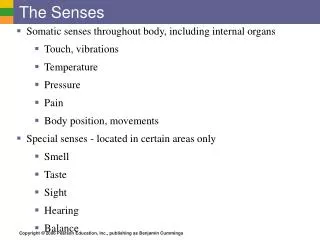
CH 15: Special Senses – The Eye. Section 1: Intro to the Eye (p. 548). The Eye and Vision. Vision is our dominant sense - 70% of all sensory receptors in the body found in eye - nearly half of cerebral cortex involved in visual processing Basic description

E N D
CH 15: Special Senses – The Eye Section 1: Intro to the Eye(p. 548)
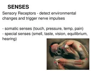
The Eye and Vision Vision is our dominant sense - 70% of all sensoryreceptors in the body found in eye - nearly half of cerebral cortex involved in visual processing Basic description - sphere w/ diameter of 2.5cm (1”) - mostly protected by cushion of fat & walls of bony orbit
CH 15: Special Senses – The Eye Section 2: Accessory Structures of the Eye (pp. 548-551)
Accessory Structures of the Eye Help to protect eye & aid in function - eyebrows - eyelids - conjunctiva - lacrimal apparatus - extrinsic eye muscles
Accessory Structures of the Eye Eyebrows - help shade eyes from excessive sunlight - prevent forehead perspiration from reaching eyes And everyone’s favorite… Some find them pointless… YIKES!! Eh…it’s only a phase…
Accessory Structures of the Eye Eyelids - protect anterior surface of eye - lids meet on sides at medial & lateral commissures “Blinking reflex” - eyelashes in follicles lined w/ very sensitive nerve endings initiate blinking reflex - occurs every 3-7 seconds - protects the eye from small particles & drying out
Accessory Structures of the Eye Eyelids - protect anterior surface of eye - lids meet on sides at medial & lateral commissures Eyelid glands- help clean/lubricate the eye & prevent irritation 1) Meibomian (tarsal) glands - open at edge of eyelid just behind eyelashes - secrete oily substance that prevents eyelid from sticking to eye & eyelashes from sticking together 2) Ciliary glands - found in eyelash follicles…modified sweat glands
Accessory Structures of the Eye Eyelid infections 1) Chalazion - infected Meibomian gland - causes painful swelling or cyst on eyelid 2) Sty - inflammation of ciliary glands - looks like small pimple around eyelash follicles
Accessory Structures of the Eye Conjunctiva - transparent membrane lining eyelids & surface of eye - only covers white part of eyes…not cornea (clear) - major function = production of lubricating mucus to help prevent dry eyes Common problems 1) Conjunctivitis - red, irritation caused by inflammation of conjunctiva 2) Pinkeye - highly contagious bacterial or viral infection of conjunctiva
Accessory Structures of the Eye Lacrimal (“tear”) apparatus - includes the lacrimal glands & ducts that drain excess tears into nasal cavity Main structures - lacrimal gland - lacrimal puncta - lacrimal sac - nasolacrimal duct
Accessory Structures of the Eye Tears - dilute saline solution containing mucus, antibodies & lysozymes to kill bacteria - tears formed by lacrimal gland located above lateral side of eye - blinking spreads tears across eye toward medial commissure - two small openings (called “lacrimalpuncta”) located on medial commissure collect tears & drain into nasolacrimal duct - nasolacrimal duct drains into nasalcavity Function of tears - wash away or dilute irritating substances - importance of emotionally induced tears is poorly understood
Accessory Structures of the Eye Extrinsic eye muscles - six strap-like muscles that control eye movements - also help maintain shape of eye - four are rectangular shaped & named for movements they allow - two are wrapped around the eye…one top & one bottom
Accessory Structures of the Eye Extrinsic eye muscles are among the most preciselycontrolled muscles in the body!
CH 15: Special Senses – The Eye Section 3: Structure of the Eyeball (pp. 551-556)
Structure of the Eyeball Wall of the eyeball contains three layers - Fibrous layer - Vascular layer - Sensory layer Internal cavity filled w/ fluids called “humors” Lens separates internal cavity into anterior & posterior segments
Fibrous Layer Fibrous layer - outermost layer of eye - composed of dense, avascular connective tissue
Fibrous Layer Two regions: 1) Sclera - white & opaque posterior region - protects & shapes eye - anchors extrinsic eye muscles 2) Cornea - transparent anterior 1/6 of eye - bends light as it enters eye - well supplied w/ pain receptors that promote blinking & tear formation when touched
Vascular Layer Vascular layer - middle, pigmented layer also known as the “uvea” Composed of three regions: 1) Choroid region - located on back portion of eye - supplies blood to all layers of eye - contains brown pigment to absorb light so it can’t be reflected w/in eye
Vascular Layer Vascular layer - middle, pigmented layer also known as the “uvea” Composed of three regions: 2) Ciliary body - ring of C.T. & muscles surrounding lens - suspends lens in position & controls its shape 3) Iris - colored part of eye - surrounds pupil (central opening that regulates entering light)
Vascular Layer More on the pupil… 1) Pupils constrict - to limit amount of light entering - in response to boredom - when looking at something repulsive 2) Pupils dilate (open) - to increase light allowed in - when looking at something appealing - in response to fear - when problem-solving
Vascular Layer More on the iris… - brown is the only pigment color found in an iris - large amounts of brown pigments create brown/black eyes - small amounts of brown pigment cause light waves to scatter resulting in blue, green, or gray eyes - newborns have blue or gray eyes…pigment develops later
Sensory Layer Sensory layer - known as the “retina” - contains millions of photoreceptors that convert light energy into a signal that can be sent to the brain
Sensory Layer Types of photoreceptors: 1) Rods - dim-light & peripheral vision receptors - verysensitive to light helping you see in the dark - do not provide sharp images (this is why objects are fuzzy when in dim lighting) 2) Cones - operate in bright light - provide color vision
Sensory Layer Other important features of retina: 1) Optic disc - known as the “blindspot” b/c it lacks photoreceptors - site where optic nerve leaves the eye 2) Maculalutea - oval region on the back of the eye - located right in the area where the lensfocuseslight 3) Fovea centralis - small spot in center of macula that contains only cones
Sensory Layer More on Fovea centralis: - due to density of cones in this spot, anything needing to be viewed critically must be focused here - size of the head of a pin, so only a very small portion of the field of view can be focused on at a given moment - explains why rapidly changing scenes (watching a train pass by, etc.) requires eyes to flickrapidly to keep image focused here
Internal Chambers & Fluids Eye divided into two segments: - chambers divided by lens & ciliary body Posterior segment - filled with clear gel called vitreous humor - gel transmits light, supports lens, provides intraocular pressure - gel forms during development & lasts lifetime Anterior segment - filled clear fluid called aqueous humor - fluid forms & drains continually & is in constant motion - supplies nutrients & oxygen to the lens & cornea - drains from eye through Canal of Schlemm
Lens Lens: - biconvex ( ), transparent, flexible, elastic, & avascular - allows precise focusing of light on retina - becomes dense, more convex, & less elastic w/ age
CH 15: Special Senses – The Eye Section 4: Physiology of Vision (pp. 556-559)
Light Electromagnetic radiation: - all energy waves (radio waves, gamma rays, X rays, etc.) Visible light - the very small portion of the electromagnetic spectrum that can stimulate our photoreceptors - can be thought of as energy packets called photons - rods & cones react to different wavelengths in visible spectrum - color seen is color being reflected off of objects - “white” objects reflect all color wavelengths - “black” objects absorb all color wavelengths
Refraction & Lenses Refraction: - bending of light caused by curvature of lens Pathway of light entering the eye: - cornea, aqueous humor, lens, vitreous humor, photoreceptors - changing shape of lens ultimately affects where light is focused - if lens is healthy, light will be directed right at foveacentralis
Focusing for Distance Vision “Far point of vision”: - the distance beyond which nochange in lens shape is needed for focusing - 20 feet away for emmetropic (“normal”) eye - means that the lens is completely relaxed when looking at something 20ft away
Focusing for Close-up Vision Requirements for focusing on things closer than 20ft: - lens shape must be changed to bend light accordingly 1) Accommodation - lens has to bulge to force light to bend more “Near point of vision” - closest point on which we can focus - determined on how much lens can bulge - in young adults = 4 inches from eye - increases w/ age…can be arms length in elderly
Focusing for Close-up Vision Requirements for focusing on things closer than 20ft: - lens shape must be changed to bend light accordingly 2) Constriction of pupils - reducing size of pupil limits extralight from entering eye - excess light would scatter inside eye causing blurriness
Focusing for Close-up Vision Requirements for focusing on things closer than 20ft: - lens shape must be changed to bend light accordingly 3) Convergence of eyes - medial rotation of eyes so that each is directed at object - closer the object, the greater degree of convergence needed *Long periods of reading or other close work require continuous accommodation, constriction, and convergence. This leads to tired eye muscles & can result in eyestrain. Periodic staring into the distance helps reduce both.
Problems with Refraction Myopia: - “near-sighted” - image is focused infront of retina - able to see objects upclose w/o problem - distant object appear blurred - usually results from an eyeball that is too long
Problems with Refraction Hyperopia: - “far-sighted” - image is focused behind retina - able to see distant object w/o problem - closeup objects appear blurred - usually results from an eyeball that is too tall
Problems with Refraction Astigmatism: - caused by unequalcurvatures in different parts of cornea/lens - require specially ground lenses, corneal implants, or laser procedures
Developmental Aspects • Vision is not fullyfunctional at birth • Babies… - born with hyperopia - see only gray tones - uncoordinated eye movements • Age 5, depth perception & colorvision well-developed • Age 6, emmetropic eyes completely developed
Developmental Aspects • With age… - lens loses clarity - dilator muscles of pupil become less efficient - ability to see clearly drastically decreased by age 70
CH 15: Special Senses – The Eye Section 5: Homeostatic Imbalances
Homeostatic Imbalances Glaucoma: - compression of the retina & opticnerve - caused when drainage of aqueous humor is blocked - leads to build-up of fluid in the eye & increased pressure Episcleritis: - inflammation of episclera (tissue between sclera & conjunctiva) - often associated w/ other diseases in the body - often affects people who spend a lot of time outdoors in very harsh environments
Homeostatic Imbalances Cataracts: - clouding of lens - can be caused by aging, diabetes, heavy smoking, frequent exposure to sunlight
Homeostatic Imbalances Blepharitis: - inflammation of margins of the eyelids Enucleation: - surgical removal of an eyeball Exophthalmos: - anteriorly bulging eyes - often a sign of a hyperactive thyroid gland
Homeostatic Imbalances Scotoma: - a blind spot other than the normal blind spot - often indicates the presence of a brain tumor or stroke Trachoma: - highly contagious bacterial infection of cornea & conjunctiva - caused by chlamydia - ultimately causes blindness if left untreated