Download
1 / 68
911 likes | 2.35k Views
PERFORATED PEPTIC ULCER. Definition. A peptic ulcer is a mucosal defect which penetrates the muscularis mucosae and muscularis propria Produced by acid-pepsin aggression. PEPTIC ULCER DISEASE PUD.

E N D
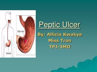
Definition • A peptic ulcer is a mucosal defect which penetrates the muscularis mucosae and muscularis propria • Produced by acid-pepsin aggression.
PEPTIC ULCER DISEASEPUD • 70-90% of ulcers are associated with Helicobacter pylori a spiral-shaped bacterium that lives in the acidic environment of the stomach. • Ulcers can also be caused or worsened by drugs such asAspirin, Plavix, NSAIDs
PEPTIC ULCER DISEASEPUD • More peptic ulcers arise in the duodenum rather than in the stomach. • About 4% of stomach ulcers are caused by a malignant tumor, so multiple biopsies are needed to exclude cancer. • Duodenal ulcers are generally benign.
CLASSIFICATION By Region/Location • Stomach • Duodenum • Esophagus • Meckel’s Diverticulum
Modified Johnson Classification of peptic ulcers • Type I: Ulcer along the body of the stomach, most often along the lesser curve at incisura angularis along the locus minoris resistentiae. • Type II: Ulcer in the body in combination with duodenal ulcers. Associated with acid oversecretion.
Modified Johnson Classification of peptic ulcers • Type III: In the pyloric channel within 3 cm of pylorus. Associated with acid oversecretion. • Type IV: Proximal gastroesophageal ulcer • Type V: Can occur throughout the stomach. Associated with chronic NSAID use .
Symptoms • Usually, children and the elderly do not develop any symptoms unless complications have arisen.
Complications • Upper digestive bleeding is the most common complication. • Sudden large bleeding can be life-threatening. • It occurs when the ulcer erodes one of the blood vessels, such as the gastroduodenal artery.
Complications • Perforation often leads to catastrophic consequences. • Erosion of the gastro-intestinal wall by the ulcer leads to spillage of stomach or intestinal content into the abdominal cavity. • Perforation at the anterior surface of the stomach leads to acute peritonitis, initially chemical and later bacterial peritonitis. The first sign is often sudden intense abdominal pain. • Posterior wall perforation leads to pancreatitis; pain in this situation often radiates to the back. • Perforation in the CBD- aerobilia, colangitis
Complications • Penetration is when the ulcer continues into adjacent organs such as the liver and pancreas. • Gastric outlet obstruction - scarring and swelling due to ulcers causes pyloric narrowing. Patient often presents with severe vomiting. • Cancer is included in the differential diagnosis (elucidated by biopsy), Helicobacter pilory as the etiological factor making it 3 to 6 times more likely to develop stomach cancer from the ulcer.
Differential diagnosis of epigastric pain • Peptic ulcer • Gastritis • Stomach cancer • Gastroesophageal reflux disease • Pancreatitis • Hepatic congestion • Cholecystitis • Biliary colic • Inferior myocardial infarction • Referred pain: pleuresy, pericarditis • Superior mesenteric artery syndrome
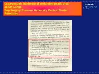
Perforated peptic ulcer • The first report of a series of patients presenting with perforation of a duodenal ulcer was made in 1817 by Travers. • The earliest operative description was made by Mikulicz in 1884 but the first successful operation for a perforated duodenal ulcer was not until 1894.
PPU patients - various treatment protocols: - an expanded role for non-operative treatment, - a developing role for laparoscopic surgery - more precise identification of those patients suitable for immediate definitive ulcer management.
Pathogenesis and epidemiologyPPU • Perforation complicates DU about 1/2 as often as bleeding • Most perforated ulcers are on the anterior surface of the duodenum. • The patient population tends to be elderly (mean age 60–70), chronically ill patients often (40–50%) taking ulcerogenic medication.
Pathogenesis and epidemiology • The fall in admissions for uncomplicated duodenal ulcers noted since the 1970's is largely attributed to the introduction of H2 antagonists. • In the third world the clinical picture is different: • a high male : female ratio (approximately 8 : 1), • younger age • a strong link with cigarette smoking. • In addition in the third world there is a high incidence of patients who present late - accounting for the high mortality (20%)
Pathogenesis and epidemiology • Helicobacter pylori is implicated in 70–92% of all PPU • The second most common cause of perforated duodenal ulcer is the ingestion of NSADs. • The least common cause is pathologic hypersecretory states, such as Zollinger-Ellison syndrome, although these should be considered in all cases of recurrent ulcer after adequate treatment.
Pathogenesis and epidemiology • In the modern treatment of PDU it must be born in mind that appropriate treatment of H. pylori infection results in eradication of the bacteria and healing of uncomplicated ulcers in more than 90% of cases. • Therefore, in the majority of cases duodenal ulcer may be regarded as a curable infectious disease or related to the ingestion of an ulcerogenic drug.
Diagnosis • The most characteristic symptom is the suddenness of the onset of epigastric pain. • The pain rapidly becomes generalised although occasionally it moves to the RLQ. • The patient stays still.
Diagnosis • There may be a history of previous dyspepsia, previous or current treatment for a DU, or ingestion of NSADs. • On examination the patient is in obvious pain. • Hypotension is a late finding as is a high fever. • The abdominal findings are characteristically described as of board-like rigidity.
Diagnosis • With time the patient may improve with dilution of the duodenal contents by exudate from the peritoneum but this is later replaced by the signs and symptoms ofbacterial peritonitis. • Once an ulcer perforates, the subsequent clinical picture is influenced by whether or not the ulcer self seals.
Diagnosis • In approximately 40–50% of cases the ulcer self-seals with omentum or by fusion of the duodenum to the underside of the liver between the gallbladder and the falciform ligament. • This is important when one considers whether or not laparotomy is indicated to deal with the perforation itself as will be seen below. • On an erect Chest X Ray free air can be seen in about 80% of cases.
Diagnosis • In doubtful cases a water-soluble gastroduodenogram will show the leak from the duodenum or its sealing. • This can be a useful test when one is considering non-operative treatment or in the situation where the diagnosis is in doubt.
What is the typical presentation in patients with perforated peptic ulcer? • Studies have shown that of patients presenting with complicated peptic ulcer disease (PUD), nearly half have no history of the disease. • In younger patients, severe abdominal pain, which may radiate to the shoulder, may be the initial presentation. • In elderly patients, signs and symptoms may be minimal.
What is the typical presentation in patients with perforated peptic ulcer? • Other reported symptoms were dyspepsia, anorexia, nausea, and vomiting. • Duration of symptoms ranged from 4 hours to 10 days.
What role do x-rays and laboratory tests play in the diagnosis of perforated peptic ulcer? • Plain x-rays of the abdomen with the patient in the upright position have been used in diagnosing perforated ulcer. However, several case series have shown that in 30% to 50% of patients, the x-ray may be negative for free air, particularly in the elderly. • Similarly, use of water-soluble contrast medium with an upper gastrointestinal tract series or computed tomography scan may increase the diagnostic yield.
How common is perforated peptic ulcer? • Thanks to effective medical management with H2 blockers and proton pump inhibitors and the eradication of H. pylori, the incidence of PUD and the hospitalization rate for treatment have decreased. • However, the rate of complicated PUD appears to be unchanged. • For example, there was a 39.3% decrease in the hospitalization rate for PUD at Massachusetts General Hospital during the years after the introduction of cimetidine. • During that same period of time, however, the incidence of perforated peptic ulcer stayed the same.
Risk Stratification • Mortality from PPU is dependant upon the presence or absence of several risk factors. • Individual risk can also be assessed by use of APACHE II. Overall mortality is approximately 10% in most studies. • Those in whom the diagnosis is overlooked almost always die. • Risk factors affecting prognosis are: • delayed treatment (> 24 hours), • preoperative shock (BP < 100 mmHg), • concurrent serious medical illness. • When all three are present the mortality rises to 100%.
What are the risk factors for PUD and perforation? • In recent years, patients presenting with perforated PUD have tended to be: • elderly, • chronically ill, • taking one or more ulcerogenic drugs.
What are the potential complications of perforated peptic ulcer? • In most cases of perforation, gastric and duodenal content leaks into the peritoneum. • This content includes gastric and duodenal secretions, bile, ingested food, and swallowed bacteria. • The leakage results in peritonitis, with an increased risk of infection and abscess formation. • Subsequent third-spacing of fluid in the peritoneal cavity due to perforation and peritonitis leads to inadequate circulatory volume, hypotension, and decreased urine output. • In more severe cases, shock may develop.
What are the potential complications of perforated peptic ulcer? • Abdominal distension as a result of peritonitis and subsequent ileus may interfere with diaphragmatic movement, impairing expansion of the lung bases. • Eventually, atelectasis develops, which may compromise oxygenation of the blood, particularly in patients with coexisting lung disease.
Non surgical treatment • In 1935 Wangensteen noted that ulcers are able to self seal and reported on seven cases treated without surgery. • In 1946 this observation was confirmed by Taylor and he treated 28 cases without surgery with good success. • This was in the context of the high mortality and morbidity associated with surgical management at the time.
Non surgical treatment • Subsequent work showed that a water-soluble contrast study could confidently demonstrate the presence of self-sealing in 40–50% of cases. • In 1989 a trial from Hong Kong by Crofts et al. showed that non-operative treatment for PPU was accompanied by a low mortality rate and was not associated with a large number of complications when the gastroduodenogram documented a sealed perforation .
In practical terms, when the diagnosis of a perforated duodenal ulcer is established the patient is aggressively resuscitated, nasogastric suction begun, and broad spectrum antibiotic cover instituted. • If a tension pneumoperitoneum embarrasses respiration this can be aspirated to release the pneumoperitoneum. • A gastroduodenogram is performed to confirm self-sealing. • The peritonitis should resolve in 4 to 6 hours and if there is continued major fluid loss after this time or if there are progressive signs of peritonitis or increasing pneumoperitoneum then surgical intervention is required
Non surgical treatment • In the study by Crofts they carefully selected patients under the age of 70 who were haemodynamically stable, had been perforated for less than 24 hours, and could be closely monitored. • Using these criteria 70% of patients were selected to be randomized to be treated in this way. • Despite this encouraging report, and a number of other non-randomised studies, non-operative treatment of PPU has not become popular. • Non-operative management has not been widely adopted probably due to problems with repeated review by senior surgeons, problems with misdiagnosis, and the lack of opportunity to perform definitive ulcer surgery.
Laparoscopic Surgery • The traditional management of a perforated duodenal ulcer has been a Graham Omental Patch and a thorough abdominal lavage. • More recently this has been shown to be able to performed using a laparoscope. The only proven advantage of the laparoscopic technique appears to be decreased postoperative pain. • Operating times are longer compared to open techniques and hospital time appears to be similar to conventional treatment. • This technique has not been subjected to any large prospective trials and at present must not be considered as standard management.
Immediate Definitive Surgery • Over the last one hundred years a number of attempts have been made to improve upon the results of simple closure and lavage. • This has been in response to the large number of patients (25–85%) who continue to have symptoms attributable to their ulcer diathesis after surgery. • The incidence of ulcer symptoms, in most studies, is related to whether the ulcer is acute or chronic (history greater than 3 months) as judged by preoperative history. • Patients with chronic ulcer symptoms generally have a higher incidence of subsequent recurrent ulcers. Up to 71% come to require subsequent definitive surgery although in most studies the figure is considerably less than this.
Pathogenic surgery • There is good evidence that, in the emergency situation, highly selective vagotomy (proximal gastric, or parietal cell vagotomy) combined with simple omental patch closure of the perforation, in patients without the risk factors mentioned above, is just as effective as that performed in the elective setting (Grade C). • This is associated with a less than 1% mortality rate and a 4–11% ulcer recurrence rate. The success of this operation is surgeon-dependent. • Truncal vagotomy with drainage has its advocates as an expedient operation familiar to most surgeons.
Immediate Definitive Surgery • Immediate definitive ulcer surgery has not gained widespread popularity due to an unfounded feeling that it is associated with a higher mortality than simple closure. • Many agree that an appropriate approach is to select only those with a chronic history (> 3 months) and without preoperative risk factors for immediate definitive surgery. • A major difficulty is defining preoperatively the patients who truly have a chronic ulcer history as many patients are too unwell to give a reliable history. • In addition, many ulcers have a silent ulcer history with as many as 70% of perforations occurring as the first manifestation of the ulcer diathesis.
It must be recalled, however, that in the developed world the surgeon's major role in the management of PPU will continue to be the performance of lifesaving operations in elderly unfit patients
Conservative treatment • There has been a return to the use of simple omental patch closure since the late 1970's with the introduction of post-operative H2 antagonists and more recently Proton Pump Blockers. • Over the last 10 years this trend has only grown stronger due to the discovery of the role of H. pylori in the pathogenesis of duodenal ulcer. • Given that H. pylori is able to be implicated in up to 90% of perforated duodenal ulcers it would seem logical to utilise patch closure and subsequent antibiotic treatment of the infectious agent saving definitive surgical ulcer management for those who fail this regimen. This has recently been tested in a randomiced controlled trial from Hong Kong where it was found that simple dosure of duoderal ulcer perforation with eradication of H. pylori resulted in ulcer healing in 78% of patients with only a 48% recurrence rate at one year (Grade A).
Conservative treatment • The counter argument is that PPU patients represent a subgroup of patients with a very vigorous ulcer diathesis and simple closure of the ulcer and treatment of the infecting organisms may not be adequate. • Until further clinical trials are performed in relevant population groups we will not know the definitve answer.
Therapy for H. pylori • The most effective present first line regimen for eradicating H. pylori, with approximately 90% eradication rates, is a combination of Bismuth, Metronidazole and Amoxycillin or Tetracycline given for a period of 14 days with or without proton pumpblocker (Grade A/B). • It is important to check for eradication, as resistance is developing to the first line regimens, especially in the third world. Tests to confirm eradication include endoscopy and biopsy and non invasive tests such as serology or breath testing for urea. • At present it would seem prudent to perform a post-operative endoscopy to confirm H. pylori eradication, confirm the diagnosis, and to check ulcer healing. • If H. pylori is eradicated the risk of reinfection is low, in the order of 1% per year, and the ulcer is likely to heal. If the ulcer heals there is no role for continuing with antisec-retory drugs. If the patient needs to continue Non Steroidal Antiinflammatories the most effective prophylactic drug is Misoprostol (Grade B).
Treatment • The present management of perforated duodenal ulcer is in flux. The great debates of earlier in the century regarding simple patch closure versus immediate definitive surgery have been complicated by the arguments for and against laparotomy, the introduction of laparoscopy, and the discovery of the role of H. pylori. • Faced with a patient with a perforated duodenal ulcer the surgeon should bear in mind the role of non-operative treatment in the first instance. If this option is selected the patient will require close vigilance and a readiness to intervene at any moment that the patient shows signs of deterioration or failure to progress satisfactorily (Grade A). • If operative management is considered to be indicated, the evidence at present supports simple omental patch closure and lavage followed by antibiotic treatment for H. pylori (Grade A). If the patient remains with an ulcer after surgery and H. pylori eradication then a Highly Selective Vagotomy should be performed after exclusion of a pathological hypersecretory state (Grade C).
Treatment • In the rare case of a patient who has been investigated and found to be negative for H. pylori , or who has been treated and then perforated, immediate definitive ulcer surgery should be performed in the absence of preoperative risk factors. If the surgeon is not experienced with Highly selective vagotomy then in the emergency situation a Truncal Vagotomy and Pyloroplasty is adequate treatment. If the patient perforates while taking ulcerogenic drugs a simple closure and lavage should suffice. • As the ulcer diathesis in many patients is silent, ulcer healing and H. pylori eradication should be confirmed by endoscopy. • Until a randomised prospective trial is performed the relative merits of the treatment strategies outlined above will continue to be controversial.
In one series of cases reported by Werbin, a 50% mortality rate was found in patients over age 70 with acute perforation of a duodenal ulcer who presented more than 24 hours after onset of symptoms. In this same series, patients who presented early and were operated on within 24 hours of onset of symptoms had 0% mortality. • In elderly patients with perforation, the ratio of female patients is higher. A study by Kubler and colleagues found that 57% of patients age 60 and older with perforated peptic ulcer were women. In this same study, 89% of patients presented with perforated duodenal ulcer.
What are the treatment options for perforated peptic ulcer? • Principles of conservative treatment include nasogastric suction, pain control, antiulcer medication, and antibiotics. Nonsurgical treatment has been recognized for a long time. The first major series was published by Taylor nearly 50 years ago; it reported a mortality rate of 11% in the nonsurgical treatment group, compared to 20% in the surgical group. Since then, because of improvements in operative and postoperative care, the mortality rate with surgical treatment of perforated peptic ulcer has decreased to about 5%. • Croft and colleagues compared surgical and nonsurgical treatment of perforated peptic ulcer in a randomized trial and concluded that an initial period of nonoperative treatment may be prudent except in patients over age 70. Careful observation is necessary during this period, but it may obviate the need for emergency surgery in 70% of patients. The mortality rate in this study was 5% in each group. Two-thirds of the patients over age 70 required emergency surgery.
Surgery • Several surgical techniques have been employed in the treatment of perforated peptic ulcer. • These include conservative surgery with patching of the ulcer, peritoneal lavage, and antiulcer medication, • Definitive surgery with truncal vagotomy, highly selective vagotomy, or partial gastrectomy. • Some studies have reported a high rate of ulcer recurrence in the conservative surgery group and have recommended definitive ulcer surgery for perforation.
What measures can be taken to decrease the risk of peptic ulcer disease and perforation? • Nearly one third of patients presenting with perforated peptic ulcer take NSAIDS. • Therefore, decreasing NSAID use is an important preventive measure. • For patients who must take NSAIDs, concomitant use of a proton pump inhibitor or misoprostol may decrease the risk of ulcer formation. • Smoking cessation and abstinence from alcohol should also decrease the risk of complicated PUD. • Maintaining a high index of suspicion for the disease, particularly in elderly patients, will help clinicians diagnose PUD early in its course, thus reducing morbidity and mortality.