Download
1 / 21
210 likes | 380 Views
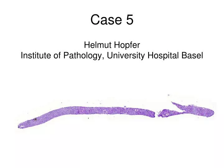
Case 5 Helmut Hopfer Institute of Pathology, University Hospital Basel. Morphological features. Intratubular crystals of needle-shaped to rectangular aggregates Brownish colour in PAS and HE stains Birefringence Von Kossa stain negative Giant cells Interstitial fibrosis and tubular atrophy.

E N D
Case 5Helmut HopferInstitute of Pathology, University Hospital Basel
Morphological features • Intratubular crystals of needle-shaped to rectangular aggregates • Brownish colour in PAS and HE stains • Birefringence • Von Kossa stain negative • Giant cells • Interstitial fibrosis and tubular atrophy
Clinical history • Subacute renal failure, serum creatinine 1250 umol/l (14.1 mg/dl) • Known autosomal dominant polycystic kidney disease since childhood • Microhematuria, minimal proteinuria • Hypertension • 2 episodes of renal colics, three and two years prior to admission, radiolucent stones on X-rays
Differential diagnosis • Nephrocalcinosis • Oxalosis • Urate nephropathy • Cystinosis • Drug-induced crystals • 2,8-Dihydroxyadenine urolithiasis
2,8-DHA urolithiasis • Autosomal recessive inherited adenine phosphoribosyltransferase deficiency (APRT), homozygosity rate 1:50'000-100'000 • Recurrent urolithiasis
♂ ♀ • APRT deficient (heterozygous) • APRT deficient (heterozygous) • ADPKD ♂ ♂ ♀ • APRT deficient (homozygous) • ADPKD • APRT deficient (heterozygous) • APRT normal • ADPKD
2,8-DHA urolithiasis Adeninemono-phosphate Adenine APRT • DNA synthesis • RNA synthesis • Energy transfer XO 8-Hydroxyadenine XO 2,8-Dihydroxyadenine APRT – Adenine phosphoribosyltransferase XO – Xanthine oxydase
2,8-DHA urolithiasis Adeninemono-phosphate Adenine APRT mutation XO 8-Hydroxyadenine XO • Excretion into the urine • Formation of crystals at physiological pH • Urolithiasis 2,8-Dihydroxyadenine APRT – Adenine phosphoribosyltransferase XO – Xanthine oxydase
2,8-DHA urolithiasis Adeninemono-phosphate Adenine APRT mutation XO Allopurinol 8-Hydroxyadenine XO 2,8-Dihydroxyadenine • Prevention of urolithiasis APRT – Adenine phosphoribosyltransferase XO – Xanthine oxydase
2,8-DHA urolithiasis • Treatment with allopurinol, low purine diet, high fluid intake • Clinical DD: urate nephropathy (radiolucent stones, standard chemical test does not differentiate) • Pathological DD: oxalosis (strong birefringence)
Differential diagnosis • Nephrocalcinosis • Oxalosis • Urate nephropathy • Cystinosis • Drug-induced crystals • 2,8-Dihydroxyadenine urolithiasis
Calcium containing crystals Nephrocalcinosis Oxalosis
Shape: round to elongate, mostly rhomboid; clusters or rosette-like Location: intraluminal, below the tubular epithelium or interstitium Colour: transparent, birefringence in polarized light (H&E stain) Special stains: von Kossa black, Alizarin orange/red Oxalate nephropathy Kossa Alizarin
Urate nephropathy • Shape: needle shaped to rectangular aggregates within an amorphous matrix • Location: tophi mostly in the medulla • Colour: FFPE biopsies – mostly dissolved, alcohol-fixed biopsies – pale to deep blue, birefringence in polarized light • Special stains: von Kossa negative, Alizarin negative
Shape: brick, hexagonal, elongated or flat Colour: FFPE biopsies – mostly dissolved, alcohol-fixed biopsies – yellow, brown to sand colour, birefringence under polarized light Special stains: von Kossa negative, Alizarin negative Cystinosis
Sulfonamides Acyclovir Methotrexate Indinavir Triamterene ... Perazella MA, Am J Med 106: 459-465, 1999 Drug-induced crystals Sulfonamide crystals (1960's)
Brownish intratubular crystals Strong birefringence Giant cells Von Kossa negative → Think of 2,8-DHA urolithiasis in all cases of oxalosis! 2,8-DHA urolithiasis