Download
1 / 20
200 likes | 317 Views
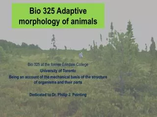
Lecture 17 Bio 325. Herbivory : (continued) Rumination Fluid feeders.

E N D
Lecture 17 Bio 325 Herbivory: (continued) Rumination Fluid feeders
Holstein cow: milk cow (left); Beef cow (right). They have been artificially selected for very different traits: basically to produce milk or to produce steaks. They have not been selected to deal with their ancestral predators. These animals are descendants of animals with claws who evolved hooves in association with a grazing lifestyle
Grasslands • Savannahs and treeless grasslands spread across much of the world by the end of the Miocene. The grasses that evolved on these prairies represented an important source of food to evolving mammals. • The problem with green plant tissue as a food is the plant cell walls: to obtain the cell protoplasm a herbivore must use its teeth (trituration) to break through the cellulose cell wall; eating and digesting the cellulose itself, a complex carbohydrate closely related to starch, is a problem because it is indigestible to all multicellular animals. Most multicellular animals cannot synthesize cellulases: only micro-organisms can make the necessary enzymes. And the grasses evolved other protections: building silica into the cellulose cell wall, a hard crystalline mineral that abraded teeth quickly.
High-crowned teeth • Cheek teeth consist of premolars and molars that function in mastication: which means they reduce food to a pulp by crushing and sliding. Watch a cow chew from the front and the lower jaw desribes a small circle, pushing lower teeth up against the upper and sliding them laterally relative to each other. [Mastication is not the same thing as maceration: to soften and separate food parts by the addition of a solvent, e.g., saliva; saliva is of course added in the mouth.] In green-plant herbivores the molars and premolars are prominent, not very differentiated, and numerous. (In a carnivore the homologous premolar cheek teeth become specialized: e.g., carnassials, for cracking bones or shearing tendons.) • The problem with green-plant tissue as food is cellulose, the material that is the basis of plant cell walls.High-crowned molars (hypsodont teeth) evolved to deal with cellulose plant walls. The part of the tooth that protrudes through the gum is much higher than teeth of nonruminants. Some grass species evolved silica that abraded teeth and produced wear (a plant defense). Teeth are comprised of cement, dentine and surface enamel, materials of differing hardness causing uneven wear. As the upper and lower jaw molars grind against each other they retain enamel cutting ridges that project from the tooth top.
Ruminantia are a suborder of the Order Artiodactyla: even-toed ungulates.Artiodactyls include about 180 species: pigs, camels, hippos, deer, giraffes, sheep, cattle, antelopes, goats, bison and others. The ruminants per se include deer, giraffes, sheep, cattle, antelopes, goats, bison, okapis.Perissodactyla are odd-toed ungulates: horses, zebras, tapirs, rhinos.Guts are muscle-invested tubes modified into a sequence of chambers, separated by valves, the walls capable of active movement [churning, peristalsis]; guts often have diverticulae (side-branches) ending blindly. The rumen and reticulum are a very large blindly ending sidebranch of the cow gut; the rumen is separated from the reticulum by a low fold, the rumino-reticuluar fold.Food is often ingested by ruminants without chewing, going down the oesophagus past the cardia (entrance into the stomach) and into the rumen. Later this unchewed material moves forward into the reticulum and there is formed into a bolus by its rough walls. The cow regurgitates this bolus up into its mouth and chews: triturates.
Tripe: lining of the reticulum, criss-crossing ridges and pits, functions in forming the grass/hay boluses that return to the mouth for further chewing; see the oesophageal groove. ideasinfood.com rumenHealth.com The oesophageal groove functions as a shunt. Reswallowed food material can reach the omasum via this groove. The groove is formed by two heavy, muscular lips/folds in the reticulum wall; these can close to create a passage and this redirects the food to the reticulo-omasal opening. If the lips remain open the material goes right into the rumen. College of Veterinary Medicine Univ. of Minnesota
Seen from the right side the rumen and anterior reticulum are apparent as a side-branch of the gut, lying eccentrically to the cow’s left side. The oesophageal groove runs in the wall of the reticulum and ends at an opening into the omasum. The abomasum is most like the stomach of non-ruminant animals and is situated just ahead of the pylorus (the sphincter valve that controls passage of material out of the stomach and into the small intestine.
Fistula: opening in the animal’s side giving access to rumen contents for study Cooperative extension system
Wallaby: cud-chewing ruminant and a marsupial that evolved its rumination separately from the artiodactyls: like them though, green plant tissue, eaten in haste, is regurgitated in leisure and masticated (chewed) further. National Geographic
Similar stomach chamber features, including an oesophageal groove evolved in wallabies independently of the evolutionary events that led to the rumen of a cow.
Homoplasy [convergence] independent adaptation resulting from similar selection processes See the section (above left re the stomach diagram) which illustrates the basis of the oesophageal groove of a wallaby. When the muscular folds are drawn together a separate tube is created which permits chewed material to be reswallowed and to pass through chamber I and directly into chamber II.
Some animals have a chamber of micro-organisms for breaking down vegetable matter that occurs later in the digestive tract: horses and rabbits use a modified caecum, part of the large intestine. Since this occurs after the absorptive region of the small intestine the material must be eaten: caecotrophy is when food is passed twice through the gut.
Langurs are monkeys that feed on the leaves of trees. They have an anterior gut chamber that houses bacteria that can break down cellulose of the plant walls. So they are adapted to feed upon green-plant tissue. Lysozymeswere evolved by mammals to deal with bacteria (as pathological body invaders) . (For example lysozymes would attack bacteria entering the eye.) Lysozymes evolved to prevent infection from bacteria. In the gut lumen there was always a need, even in mammals that did not try to exploit cellulose as food, to keep the bacterial fauna under control. The primitive lysozymes of mammals that occur in their gut and ‘handle’ the bacteria there are however, not found in their stomach. The stomach is too low in pH for these lysozymes and it contains the enzyme pepsin which would break down normal (protein) lysozymes. In the course of their evolution ruminant animals evolved special lysozymes that are resistant to acidic conditions and resistant to the presence of protein-destroying pepsin. The langur monkeys show convergence in the sequence of amino acids making up the lysozymes of their stomach with those of animals such as a cow or a moose. LYSOZYMES IN THE STOMACH OF TWO DISTANTLY RELATED TAXA SHOW AMINO-ACID SEQUENCE CONVERGENCE Reading: Stewart C-B. et al. 1987. Nature 330: 401-
Adaptations make sense only in the context of the lives of the animals that have them. This is how one ‘explains’ and understands adaptations. And many structures on an animal are affiliated in adaptation toward the same ends. The ungulate animals that evolved their stomach into chambers that house ciliate protozoa , yeasts and other micro-organisms, that use the enzyme systems of these unicellular creatures to digest the cell walls of grasses, also have high-crowned grinding teeth to successfully chew cellulose cell walls. And they have cursorialadapatations: long legs, muscles and bones and hooves, adapted for fast running: limb structures selected for distance advantage imparted to hooves. They have good depth vision and high sensitivity to objects moving at a great distance in their field of view. Knowing that such animals evolved in a prairie/grassland habitat to exploit grass as food allows us to understand how adaptations for cursorial limbs could affiliate with adaptations such as their oesophageal grooves. Ungulates adapted to exploit grass as food and to run from pursuing predators in open spaces where there is nowhere to hide evolved both rumens and long legs. Grooves and hooves were selected in the same historical context of feeding upon grasses. Something to ruminate on. Significance of ruminant adaptation
Trapping food; filtering the air; stabilimentum • Argiope(garden spider). Many spider species make stabilimenta. • These structures are thick silk radiated at the web hub and before the viscid spiral zone. • Hypotheses of selective advantage • Reinforcement; tension adjustment • Camouflage • Visual marker • Experiment showed that the presence of visual markers significantly affected the time a web could last without mechanical damage from things such as flying birds. • Eisner T. 1983. Spider web protection through visual advertisements: role of the stabilimentum Science 185-187.
Aphid feeding Rosy apple aphid P.G. Mutalik A narrow region of the face called the clypeus is, in these two insects, very different. Both insects feed on plant sap and both insert elongate stylet mouthparts into the sap tissues (phloem bundles, xylem). But the cicada has its clypeus hugely swollen to accommodate internally the dilator muscles of a large cibarial pump. Cibarial muscles in the cicada expand the space (cibarium) defined by the proximal attachments of the mouthparts, creating a negative pressure that draws the fluid food in. There is no equivalent structure in the aphid which is feeding on a fluid that is already under pressure (phloem). If you remove the aphid and leave its mouthparts inserted, they will spout sap like a little fountain.
Comparing the homologous muscles in the grasshopper (left) and the cicada (right). The grasshopper also has a cibarium ahead of its mouth. But the capacity of the grasshopper’s cibarial muscles to dilate the cibarial space is very limited compared to that of the cicada. Insects can also utilize other regions of the gut to create a pump, e.g., a pharyngeal pump. The same principle applies. Muscles that run from the exoskeleton and insert in the gut wall which by contracting can dilate a gut cavity and so create suction through the mouthparts.
The mouthparts of insects are wonderful examples of evolving structural change. Comparing the maxillae and mandibles of a grasshopper with the stylets of an aphid: the mandibles and maxillae are pulled out into long ‘straw-like’ structures with inner grooves that meet, left and right, to create channels. One channel conveys saliva down into the plant wound and the other conveys the sap upwards into the gut.
The aphid has the problem of securing a quiet backwater in its gut for secretion of enzymes and absorption of the products of digestion. The pressure of the fluid on which it feeds is a problem as it pushes the fluid through the gut at a high rate. And the aphid’s food (sap) is very dilute so it must process copious amounts. Its gut loops back on itself and then is turned in ‘switchbacks’ overtop of the anterior midgut. This ‘filter chamber’ also functions a shunt: it allows for certain portions of the fluid to bypass the posterior midgut; through differential absorption t the two overlain gut walls filter the wanted out of the unwanted.