Download
1 / 24
250 likes | 399 Views
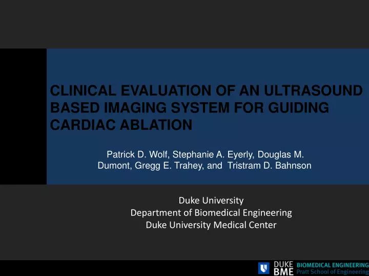
CLINICAL EVALUATION OF AN ULTRASOUND BASED IMAGING SYSTEM FOR GUIDING CARDIAC ABLATION. Patrick D. Wolf, Stephanie A. Eyerly , Douglas M. Dumont, Gregg E. Trahey , and Tristram D. Bahnson. Duke University Department of Biomedical Engineering Duke University Medical Center. Introduction.

E N D
CLINICAL EVALUATION OF AN ULTRASOUND BASED IMAGING SYSTEM FOR GUIDING CARDIAC ABLATION Patrick D. Wolf, Stephanie A. Eyerly, Douglas M. Dumont, Gregg E. Trahey, and Tristram D. Bahnson Duke University Department of Biomedical Engineering Duke University Medical Center
Introduction Normal Sinus Rhythm (NSR) • Thousands of cardiac ablation procedures performed daily to treat tachy-arrhythmias • Increase in the number of procedures that are anatomically guided rather than electrically guided • Atrial Fibrillation • 3 M now • 7 M by 2050 Atrial Fibrillation (AF)
Introduction • Treating Arrhythmias: Transcatheter Cardiac Ablation (TCA) • Single lesions directly ablate arrhythmogenic tissue • AVNRT, WPW, MAT • Lines of lesion isolate regions (pulmonary veins) - AF http://www.heart.org/HEARTORG/Conditions/Arrhythmia/PreventionandTreatmentofArrhythmia/Ablation_UCM_301991_Article.jsp
Introduction • Radiofrequency Ablation (RFA) in TCA procedures • Lines of RFA lesions must be transmural and continuous to block conduction • Electrical reconnection occurs at unablated gaps • Blood flow around catheter tip and tip-tissue contact affect lesion formation • Lesion size not predictable from delivery parameters • Real-time image based evaluation would confirm transmurality and line contiguity RFA Catheter Unablated Gap Blood flow Endocardium Transmural lesion Transmural lesion Non-transmural lesion Epicardium
Introduction • Radiofrequency Ablation (RFA) • Young’s Modulus of RFA treated tissue In vitro • RF current heats the tissuecreating a discrete and stiff lesion volume Pernot et al. Mapping Myocardial Elasticity Changes After RF-Ablation Using Supersonic Shear Imaging. Computers in Cardiology. 2009; 36:793-796
Introduction • Acoustic Radiation Force Impulse (ARFI) Imaging • An ultrasound pulse applies radiation force F [kg/cm2·s2] over a small region at the focus[1] • absorbed power = Wabsorbed, [W/cm3] • absorption coefficient = α ~ 0.00072 [Np/cm][2] • speed of sound = c ~ 1540 or 1615±15 [m/s][3] • Temporal average intensity = I [W/cm2] • Radiation force creates μm scale tissue displacements mechanical properties • Ultrasound scan lines spatially and temporally monitor tissue response Displacement [μm] Time ARFI! t1 t3 t4 t0 t2 RF Lines Axial Response Time (t) [1] Trahey, G.E., et al., Acoustic radiation force impulse imaging of the mechanical properties of arteries: in vivo and ex vivo results. Ultrasound in Medicine & Biology, 2004. [2] Sagar, K.B., et al., Quantitative ultrasonic assessment of normal and ischaemic myocardium with an acoustic microscope: relationship to integrated backscatter. Cardiovascular research, 1990. [3] Masugata, H., et al., Relationship between myocardial tissue density measured by microgravimetry and sound speed measured by acoustic microscopy. Ultrasound in Medicine and Biology, 1999.
In vitro proof of concept 4 • ARFI for RFA Lesion Assessment (in vitro proof of concept) • AccuNav ICE catheter and Siemens Anteres scanner • RFA lesion differentiable from the surrounding myocardium in vitro by measured ARFI-induced displacements • 2D ARFI able to provide lesion assessment 2 Pathology 0 μm Axial [cm] B-Mode ARFI Lateral [cm] Eyerly et. al. In vitro assessment of ARFI imaging for cardiac visualizing RFA lesions. J Cardiovasc Electrophysiol. Vol 21, 5: 557-563.
In vivo obstacles • Three (3) major obstacles to performing this imaging in vivo: • Heart stiffens and softens during each beat • Small ARFI induced displacements must be measured in the presence of substantial gross motion • A blind search of the myocardium to find lesions is extremely difficult Eyerly et. al. In vitro assessment of ARFI imaging for cardiac visualizing RFA lesions. J Cardiovasc Electrophysiol. Vol 21, 5: 557-563.
Problem 1 Stiffness Contrast • ARFI images must acquired in diastole when there is minimal cardiac motion and there is maximum elasticity contrast between the RFA lesion and the surrounding myocardium. Right ventricular wall before and after ablation. Movie made with multi-beat synthesis using a transthoracic probe positioned on the RV epicardium. • Eyerly SA, Hsu SJ, Trahey GE, Wolf PD. In vivo differentiation of myocardial ablation lesions via a stiffness ratio with acoustic radiation force impulse imaging. Eighth International Conference on the Ultrasonic Measurements and Imaging of Tissue Elasticity. Vlissingen, Netherlands: September 2009.
Problem 1 & 2: Contrast and Motion • Trigger the scanner to start the ARFI sequence during the passive filling phase of diastole. • Maximum stiffness contrast with softened myocardium • Minimum motion related to contraction
Problem 2: Motion • Take multiple scan lines before and after the ARFI ‘push’ • Track bulk motion • Fit to a quadratic function and interpolate • Subtract bulk motion • Find ‘Peak Displacement’ • Confirm motion filter with ‘zero push’ sequences Hsu, S., Acoustic Radiation Force Impulse Imaging of Myocardial Performance, in Biomedical Engnieering2009, Duke University.
Problem 3: Find the Lesions • Imaging and Guidance Tools Used in TCA Procedures • Fluoroscopy • Visualize catheter position/orientation in the body • Can not visualize soft tissue • Intracardiac Echo (B-Mode) • Identifies anatomical structures • Assess catheter-tissue contact • Can not differentiate lesion from normal tissue http://emedicine.medscape.com/article/151907-overview http://www.touchcardiology.com/img/Image/acunav.gif
Find the Lesions • Real-time guidance for TCA procedures • Electroanatomical Mapping (EAM): CARTO (Biosense/Webster) • Magnetic fields determine the 3D location of catheters • NaviStar™mapping/ablation catheter: contains a location coil that measures magnetic field strength • All points plotted relative to a reference patch on the patient’s back http://www.ipej.org/0801/bhakta.htm http://www.pages.drexel.edu/~gn52/MEDICAL_ROBOTICS/Downloads_files/Carto%20XP%20EP%20Navigation%20System.pdf
Find the Lesions • Real-time guidance for TCA procedures • Electroanatomical Mapping (EAM): CARTO (Biosense Webster) • Construct 3D cartoon of chamber geometry • Mapping catheter collects endocardial EGs, local activation times (LAT) • Visualization of electrical propagation, identifies arrhythmogenic regions
Find the Lesions • Tracking and 2D ICE based imaging • Magnetic tracking of imaging tip • Registered display of B-mode images on cartoon • Shows “where you are looking” 64 element phased array http://www.biosensewebster.com/products/navigation/cartosound.aspx Hsu, S.J., et al., Challenges and implementation of radiation-force imaging with an intracardiac ultrasound transducer.Ieee Transactions on Ultrasonics Ferroelectrics and Frequency Control, 2007. 54(5): p. 996-1009.
In vivo testing Right atrium mapped using CARTO EAM system No Lesion • Paced from coronary sinus catheter • Mapped electrical activation using CARTO • ARFI imaged normal tissue • Created a lesion line with ≈1 cm gap • ARFI imaged line and gap • Closed the gap • ARFI imaged line • Poster by Eyerly Lesion with Gap Gap Closed
Normal and Gap Images RFA Lesion Gap Gap RFA Lesion
In vivo Results • Linear RFA • ARFI image lesion assessments correlate with electrical block in CARTO LAT maps TA μm Gap at TA RFA Lesion Continuous RFA Lesion RFA Lesion TA 81ms CARTO LAT Maps 24ms 8 6.4 4.8 3.2 1.6 0
Moving to the Clinic - ARFIi Clinical Tool • Research Tool • Acquire ECG synchronous ARFI data on S2000 scanner • Download data to laptop • Calculate and display ARFI displacements on laptop screen • Clinical Tool • Acquire ECG synchronous ARFI data on S2000 scanner • Process and display ARFI data on S2000 screen • Done by Duke in collaboration with Siemens Medical
Moving to the Clinic – FDA Considerations • Mechanical Index (MI) • FDA limit 1.9 • Measured peak for our sequences <1.6 • Face Heating • FDA limit < 6 °C in standing water • Siemens internal limit <2 °C • Measured change for our sequences <2 °C
Moving to the Clinic • Clinically, we seek to understand • The elasticity contrast for lesions in senescent and diseased hearts • The ability to access all lesions in both the right and left atria with a limited field of view • The effect of atrial fibrillation on ARFIi contrast • The correlation of ARFIi evaluation with electrical outcome in atrial flutter and atrial fibrillation ablation procedures
Clinical Study Outline • Patients undergoing AFl ablation or AF ablation • Following each round of ablation in the normal therapeutic procedure image normal and lesioned tissue with ARFI • blinded results to the physician • Characterize “normal” and ablated tissue • In sinus rhythm • In AF
Summary ARFI imaging of ablation lesions can be implemented clinically with significant but minor modifications to existing equipment commonly used in the EP lab (CARTO, ICE). Lesion boundary correlates with ARFI imaged stiffness boundary Electrical block correlates with ARFIi determined contiguous and transmural lesions The tools for clinical evaluation have been created
Acknowledgements Thank you to Dr. Kapur et al for organizing workshop NIH Grant: R21-EB-007741 NIH Grant: R01-EB-012384 Biosense Webster and Siemens Medical Solutions USA, Inc. for their hardware and system support