Download
1 / 40
410 likes | 594 Views
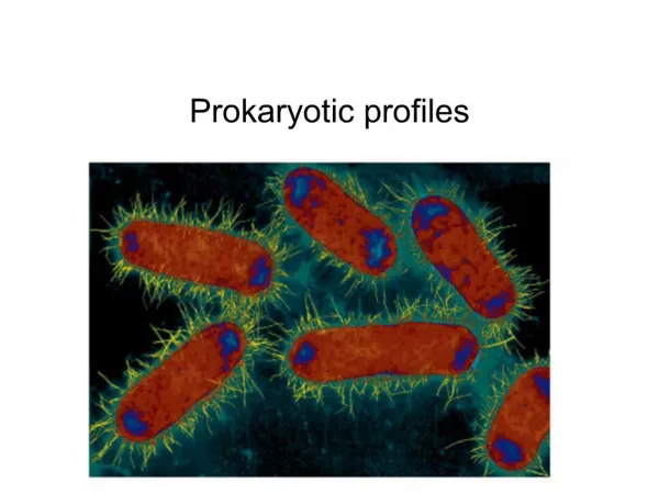
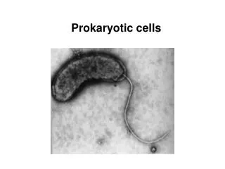
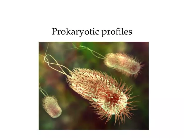
Prokaryotic profiles. When did prokaryotes first appear?. What are the basic differences?. How do prokaryotes compare in size with other microorganisms?. How do I describe their shapes? . Is a species always the same shape?. Generally, yes Monomorphic however, environment can alter

E N D
How do prokaryotes compare in size with other microorganisms?
Is a species always the same shape? • Generally, yes • Monomorphic • however, environment can alter • Some are pleiomorphic Photo from: uhavax.hartford.edu/bugl/Yersinia-pestis.jpg
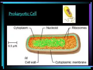
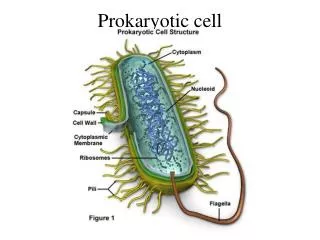
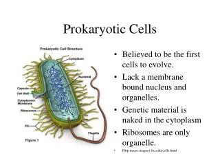
The parts Starting from the outside and working inward
What’s outside the cell wall? • Glycocalyx • Carbohydrates and/or peptides • Viscous • Can protect bacterium • Some help cells attach • Teeth: Streptococcus mutans • Capsule • Organized • Firmly attached to cell wall • Slime layer • Unorganized • Loosely attached
What are flagella? • Singular = __________ • Monotrichous • Amphitrichous • Lophotrichous • Peritrichous
What do flagella do? • Run and tumble • Swarming • Allow for taxis • Chemotaxis • Phototaxis • dancing bacteria!
What are axial filaments? • AKA periplasmic flagella • Fibril bundles that spiral around cell
What’s the difference between fimbriae and pili? • Found primarily on gram-negative bacteria • For attachment, not movement • Pilin protein • Fimbriae (fimbria, singular) • Attachment • E.g. to mucosal membranes • Pili (pilus, singular) • For DNA exchange only
What’s in the cell wall? • Peptidoglycan (PPG) • AKA murein • NAG-NAM disaccharide • NAG = N-acetyleglucosamine • NAM = N-acetylemuramic acid • Lysozyme disrupts NAG-NAM bond • If lysis doesn’t occur, cell is called a protoplast • Linked with tetrapeptide • Penicillin disrupts lysis • penicillin killing cells
What’s in the cell wall? • Gram positive bacteria • Many PPG layers • Teichoic acids • Different types • Used for antigenic specificity tests
What’s in the cell wall? • Gram negative bacteria • One or few PPG layer(s) • Outer membrane: lipopolysaccharides (LPS), lipoproteins, phospholipids • Periplasm separates LPS from the PM (PPG is in periplasm) • Provides barrier to some antibiotics, digestive enzymes • Porins allow for access into cell • LPS used for specific antigen tests to I.D. species
How does this relate to gram staining? • Hint: What does the LPS layer covering the gram negative cell do to it?
What are atypical cell walls? • Mycoplasma • Smallest known independent bacteria • No cell walls • Often mistaken for viruses • PM has sterols to prevent lysis • Mycobacteria • Mycolic acid in cell wall • Hydrophobic • Acid-fast stain identifies • Tuberculosis, leprosy • Archaea • Some have cell walls but not with PPG • Pseudo-murein Mycoplasm pneumoniae
What happens if the cell wall is damaged? • Lysozyme lyses gram positive, but usually does not harm gram negative to the same extent • Why? • Protoplast: gram positive • Spheroplast: gram negative • Osmostic lysis • If placed in a hypotonic environment
What’s inside the cell wall? • Plasma membrane • Phospholipidbilayer • Fluid mosaic theory • Segregates DNA during binary fission • Secretes enzymes to make PPG, teichoic acid • ATP production • Selective permeable membrane… • Active vs. passive transport
What kinds of passive transport exist? • Simple diffusion • For small, lipid-soluble substances • Osmosis • Water movement via diffusion • Happens whenever difference in concentration across PM • Note: water often moves because solutes can’t • Because PM is only semi-permeable Simple diffusion
What do you think will happen? Left side increases with water Right side increases with water No net movement of water Click here to show what actually happens: answer animation
What is osmosis? • Water concentration depends on number of solutes in it • Hypertonic • Isotonic • Hypotonic • Water moves down its concentration gradient until osmolarity is equal
10% glucose 20% glucose 10% glucose Distilled water What do you think will happen? For each, choose from A. No net change B. cell swells C. cell shrinks
What is facilitated passive diffusion? • Protein-assisted diffusion • Transporters or carriers • Amino acids, glucose • Channels (AKA “pores”) • Most are gated (usually closed)
What is active transport? • Movement of solute against gradient • Can you think of examples of where this might happen in your body? • Requires energy b/c moving against gradient • From ATP • Proteins sometimes called “pumps”
What is group translocation? • Type of active transport • Only in prokaryotes • Chemically altered as it is pulled across PM into cell • Once inside, cannot exit • E.g. glucose phosphorylation • Animation
What is the cytoplasm? • Eukaryotes • Cytosol + organelles • Prokaryotes: all stuff inside cell • 80% water + nuclear area, ribosomes, inclusions (storage areas)
What is the nuclear area? • AKA nucleoid • Circular, double-stranded DNA • Called bacterial chromosome • Plasmid • Also double-stranded DNA • Independent replication • Associated with PM proteins • Can gain or lose without killing cell • Can provide resistance to antibiotics, etc.
What are ribosomes? • Manufacture proteins • Two subunits • Each with proteins and rRNA • 70S ribosomes (smaller than eukaryotes) • 50S and 30S subunits • Eukaryotes = 80S ribosomes (60S + 40S)
What are inclusions? • Storage areas • Metachromatic granules • Collectively called volutin • Phosphate reserve for making ATP • Polysaccharide granules • Iodine stain shows these • Others with • Lipids, sulfur granules, etc. • Magnetosomes: iron oxide
What are endospores? • Usually gram-positive bacteria • Not a reproductive structure • Survival structure for bad times • Inside PM • Form thick walls • Tolerate high heat, dehydration, poisons, radiation • Can survive up to 25 to 40 M years!!!!!!!! • Problem for food industry! • Botulism
How are spores formed? • Sporogenesis • Usually resource scarcity triggers • Carbon, nitrogen, etc. • Spore is highly dehydrated • Vegetative state • Favorable conditions • Germination
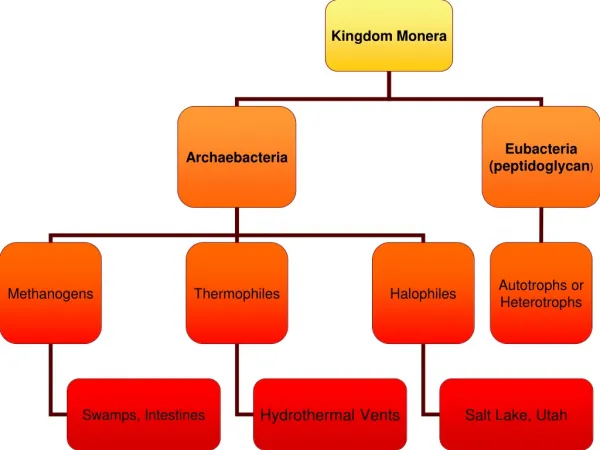
What are taxonomic groups of bacteria? • Gracilicutes • Gram - • Firmicutes • Gram + • Tenricutes • (no cell cell—e.g. mycoplasmas) • Mendosicutes • (Archaebacteria)
What’s the difference between a species and a strain? • Species: share similar pattern of traits • Subspecies/strain/type: same species with differing characteristics • Serotypes: unique antibody response in host
What about unusual bacteria? • Rickettsias • Parasitic, gram negative • Arthropod vector • RMSF • Q Fever
What about unusual bacteria? • Chlamydias • Parasitic • No vector Photo from: http://www.sexually-transmitted-diseases.info/images/std_chlamydia.jpg
What about Mendosicutes? • No PPG • 70S • Extremophiles • Halophiles • Thermophiles • Methanophiles Photo from: http://people.westminstercollege.edu/faculty/tharrison/gslfood/studentpages/pinkwater2.JPG