Download
1 / 1
20 likes | 162 Views
Detection of phorid fly DNA in honey bees through PCR . Samuel Barton, Zachary De Moulin, Mark Martinez and Tia Albertson, Kim Mogen, University of Wisconsin-River Falls, WI. Introduction

E N D
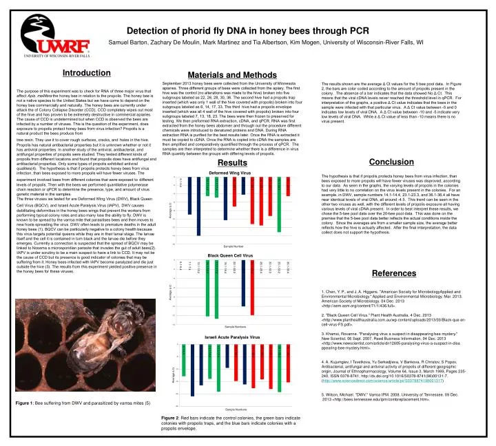
Detection of phorid fly DNA in honey bees through PCR Samuel Barton, Zachary De Moulin, Mark Martinez and Tia Albertson, Kim Mogen, University of Wisconsin-River Falls, WI Introduction The purpose of this experiment was to check for RNA of three major virus that affect Apis. melliferathe honey bee in relation to the propolis. The honey bee is not a native species to the United States but we have come to depend on the honey bee commercially and naturally. The honey bees are currently under attack the of Colony Collapse Disorder (CCD). CCD completely wipes out most of the hive and has proven to be extremely destructive in commercial apiaries. The cause of CCD is undetermined but when CCD is observed the bees are infected by a number of viruses. This is the question of the experiment, does exposure to propolis protect honey bees from virus infection? Propolis is a natural product the bees produce from tree resin. They use it to cover rough surfaces, cracks, andholes in the hive. Propolis has natural antibacterial properties but it is unknown whether or not it has antiviral properties. In another study of the antiviral, antibacterial, and antifungal properties of propolis were studied. They tested different kinds of propolis from different locations and found that propolis does have antifungal and antibacterial properties. Only some types of propolis exhibited antiviral qualities(4). The hypothesis is that if propolis protects honey bees from virus infection, than bees exposed to more propolis will have fewerviruses. The experiment involved bees from different colonies that were exposed todifferent levels of propolis. Then with the bees we performed quantitative polymerase chain reaction or qPCR to determine the presence, type, and amount of virus genetic material in the samples. The three viruses we tested for are Deformed Wing Virus (DWV), Black Queen Cell Virus (BQCV), and Israeli Acute Paralysis Virus (IAPV),DWV causes debilitating deformities in the honey bees wings that prevent the workers from performing typical colony roles and also many lose the ability to fly. DWV is known to be spread by the varroa mite that parasitises bees and then moves to new hosts spreading the virus. DWV often leads to premature deaths in the adult honey bees (1). BQCV can be particularly negative to a colony health because this virus targets potential queens while they are in their larval stage. The larvae itself and the cell it is contained in turn black and the larvae die before they emerges. Currently a connection is suspected that the spread of BQCV may be linked to Nosema a microsporidian parasite that invades the gut of adult bees(2). IAPV is under scrutiny to be a main suspect to have a link to CCD. It may not be the cause of CCD but its presence is good indicator of colonies that may be suffering from it. Honey bees infected with IAPV become paralyzed and die just outside the hive (3). The results from this experiment yielded positive presence in the honey bees for these viruses. . Materials and Methods September 2013 honey bees were collected from the University of Minnesota apiaries. Three different groups of bees were collected from the apiary. The first hive was the control (no alterations was made to the hive) broken into five subgroups labeled as 22, 26, 28, 30, 36. The second hive had a propolis trap inserted (which was only 1 wall of the hive covered with propolis) broken into four subgroups labeled as 6, 14, 17, 33. The third hive had a propolis envelope inserted (which was all 4 wall of the hive covered with propolis) broken into four subgroups labeled 7, 13, 18, 23. The bees were then frozen to preserved for testing. We then preformed RNA extraction, cDNA, and qPCR. RNA was first extracted from the honey bees abdomen and through out the procedure different chemicals were introduced to denatured proteins and DNA. During RNA extraction RNA is purified for the best results later. Once the RNA is extracted it must be copied to cDNA. Once the RNA is copied into cDNA the samples are then amplified and comparatively quantified through the process of qPCR. The samples are then interpreted to determine whether there is a difference in virus RNA quantity between the groups with differing levels of propolis. The results shown are the average Δ Ct values for the 5 bee pool data. In Figure 2, the bars are color coded according to the amount of propolis present in the colony. The absence of a bar indicates that the data showed No Δ Ct. This means that the viral cDNA levels never reached the threshold level in qPCR. For interpretation of the graphs, a positive Δ Ct value indicates that the bees in the sample were infected with that particular virus. A Δ Ct value between -5 and 0 indicates low levels of viral DNA. A Δ Ct value between -10 and -5 indicate very low levels of viral DNA. While a Δ Ct value of less than -10 means there is no virus present. Conclusion Results The hypothesis is that if propolis protects honey bees from virus infection, than bees exposed to more propolis will have fewer viruses was disproved, according to our data. As seen in the graphs, the varying levels of propolis in the colonies had very little to no correlation on the virus levels present in the colonies. For an example, in DWV, sample numbers 14.1-14.4, 23.1-23.3, and 36.1-36.4 all have near identical levels of viral DNA, all around -4.5. This trend can be seen in the other two viruses as well, with the different levels of propolis exposure all having various levels of viral cDNA present. In order to best interpret these results, we chose the 5-bee pool data over the 20-bee pool data. This was done on the premise that the 5-bee pool data better reflects the actual conditions inside the colony. Since the averages are from a smaller sample size, the average better reflects how the hive is actually affected. After the final interpretation, the data collect does not support the hypothesis. References 1. Chen, Y. P., and J. A. Higgens. "American Society for MicrobiologyApplied and Environmental Microbiology." Applied and Environmental Microbiology. Mar. 2013. American Society of Microbiology. 04 Dec. 2013 <http://aem.asm.org/content/71/1/436.full>. 2. "Black Queen Cell Virus." Plant Health Australia. 4 Dec. 2013 <http://www.planthealthaustralia.com.au/wp-content/uploads/2013/03/Black-que en-cell-virus-FS.pdf>. 3. Khamsi, Roxanne. "Paralysing virus a suspect in disappearing bee mystery." New Scientist. 06 Sept. 2007. Reed Business Information. 04 Dec. 2013 <http://www.newscientist.com/article/dn12605-paralysing-virus-a-suspect-in-disa ppearing-bee-mystery.html>. 4. A. Kujumgiev, I Tsvetkova, Yu Serkedjieva, V Bankova, R Christov, S Popov, Antibacterial, antifungal and antiviral activity of propolis of different geographic origin, Journal of Ethnopharmacology, Volume 64, Issue 3, March 1999, Pages 235-240, ISSN 0378-8741, http://dx.doi.org/10.1016/S0378-8741(98)00131-7. (http://www.sciencedirect.com/science/article/pii/S0378874198001317) 5. Wilson, Michael. "DWV." Varroa IPM. 2008. University of Tennessee. 09 Dec. 2013 <http://bees.tennessee.edu/ipm/combreplacement.htm>. Figure 1: Bee suffering from DWV and parasitized by varroa mites (5) Figure 2: Red bars indicate the control colonies, the green bars indicate colonies with propolis traps, and the blue bars indicate colonies with a propolis envelope.