Download
1 / 2
20 likes | 142 Views
Supplementary figure 1. Transmission electron micrograph of a negatively stained cell of strain S1-08 T . Bar, 1 μm. Supplementary figure 2. Thin-layer chromatograms showing the total polar lipid compositions of (a) S1-05 and (b) S1-08 T .

E N D
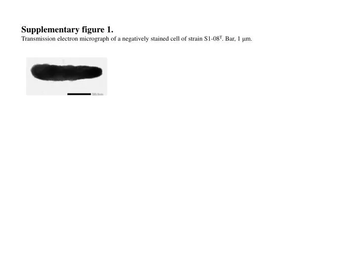
Supplementary figure 1. Transmission electron micrograph of a negatively stained cell of strain S1-08T. Bar, 1 μm.
Supplementary figure 2. Thin-layer chromatograms showing the total polar lipid compositions of (a) S1-05 and (b) S1-08T. Total polar lipids were detected by spraying the plate with 5 % molybdophosphoric acid. AL, unidentified aminolipid; GL, unidentified glycolipid; PE, phosphatidylethanolamine; L1, unidentified polar lipids. a) S1-05 L1 b) S1-08T GL1 GL2 S2 AL1 L1 PE AL2 GL1 S1 S2 GL2 AL1 PE AL2 S1