Download
1 / 116
1.16k likes | 2.32k Views
Lesson 1 - Types of Muscles - Characteristics. The Muscular System. The Muscular System. Muscles are responsible for all types of body movement Three basic muscle types are found in the body Skeletal muscle Cardiac muscle Smooth muscle. Characteristics of Muscles.

E N D
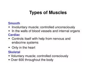
The Muscular System • Muscles are responsible for all types of body movement • Three basic muscle types are found in the body • Skeletal muscle • Cardiac muscle • Smooth muscle
Characteristics of Muscles • Skeletal and smooth muscle cells are elongated (muscle cell = muscle fiber) • Contraction of muscles is due to the movement of microfilaments • All muscles share some terminology • Prefixes myo and mys refer to “muscle” • Prefix sarco refers to “flesh”
Comparison of Skeletal, Cardiac, and Smooth Muscles Table 6.1 (1 of 2)
Comparison of Skeletal, Cardiac, and Smooth Muscles Table 6.1 (2 of 2)
Anatomical (structural) differences (possible answers may include) Cell shape and appearance • Skeletal muscles are multinucleate while smooth and cardiac muscles are uninucleate • Skeletal & cardiac muscles have striations while smooth do not • Only cardiac muscle has intercalated discs Location • Skeletal muscle is attached to bones, cardiac muscle is in the heart, smooth muscle lines the walls of hollow organs (ex. Digestive tract)
Physiological (functional) differences (possible answers may include) Regulation of contraction • Skeletal muscle is under voluntary control while smooth and cardiac muscle are under involuntary control Speed of contraction • Skeletal muscles can be slow or fast to contract but cardiac muscles contract slow & smooth muscles contract very slow. Rhythm of contraction • Cardiac and some smooth muscles have rhythmic contraction, skeletal does not
Smooth Muscle Characteristics Figure 6.2a • Lacks striations • Spindle-shaped cells • Uninucleate • Involuntary— no conscious control • Found mainly in the walls of hollow organs
Cardiac Muscle Characteristics Figure 6.2b • Striations • Usually uninucleate • Branching cells • Joined to another muscle cell at an intercalated disc • Involuntary – no conscious control • Found only in the heart
Skeletal Muscle Characteristics • Most are attached by tendons to bones • Cells are multinucleate • Striated — have visible banding • Voluntary — subject to conscious control
Skeletal Muscle Attachments • Epimysium blends into a connective tissue attachment • Tendons—cord-likestructures that connect muscle to bone • Mostly collagen fibers • Often cross a joint due to toughness and small size • Aponeuroses—sheet-likestructures • Attach muscles indirectly to bones, cartilages, or connective tissue coverings
Skeletal Muscle Attachments • Sites of muscle attachment • Bones • Cartilages • Connective tissue coverings
Skeletal Muscle Functions • Produce movement • Maintain posture • Stabilize joints • Generate heat
Lesson 2- Skeletal Muscle Structure- connective tissue wrappings
Connective Tissue Wrappings of Skeletal Muscle • Cells are surrounded and bundled by connective tissue • Endomysium — encloses a single muscle fiber • Perimysium — wraps around a fascicle (bundle of muscle fibers) • Epimysium — covers the entire skeletal muscle • Fascia — on the outside of the epimysium
Lesson 3- Muscle Stimulation- stimulation & contraction - nerve stimulus & action potential - transmission of impulse to muscle - sliding filament theory
Stimulation and Contraction of Single Skeletal Muscle Cells • Excitability (also called responsiveness or irritability)—ability to receive and respond to a stimulus • Contractility—ability to shorten when an adequate stimulus is received • Extensibility—ability of muscle cells to be stretched • Elasticity—ability to recoil and resume restinglength after stretching
The Nerve Stimulus and Action Potential • Skeletal muscles must be stimulated by a motor neuron (nerve cell) to contract • Motor unit— one motor neuron and all the skeletal muscle cells stimulated by that neuron
The Nerve Stimulus and Action Potential Figure 6.4a
The Nerve Stimulus and Action Potential Figure 6.4b
The Nerve Stimulus and Action Potential • Neuromuscular junction • Association site of axon terminal of the motor neuron and muscle • Synaptic cleft • Gap between nerve and muscle • Nerve and muscle do not make contact • Area between nerve and muscle is filled with interstitial fluid
The Nerve Stimulus and Action Potential Figure 6.5a
The Nerve Stimulus and Action Potential Figure 6.5b Synaptic cleft
Transmission of Nerve Impulse to Muscle • Neurotransmitter—chemicalreleased by nerve upon arrival of nerve impulse • Carries the impulse across the synaptic cleft • The neurotransmitter for skeletal muscle is acetylcholine (ACh) • Acetylcholine attaches to receptors on the sarcolemma of the muscle cells • Sarcolemma becomes permeable to sodium (Na+)
Transmission of Nerve Impulse to Muscle Figure 6.5c • Sodium rushes into the cell generating an action potential • Once started, muscle contraction cannot be stopped
Transmission of Nerve Impulse to Muscle Figure 6.6
The Sliding Filament Theory of Muscle Contraction • Activation by nerve causes myosin heads (cross bridges) to attach to binding sites on the thin filament; requires energy in form of ATP • Myosin heads then pull thin filaments toward the center of the sarcomere • This continued action causes a sliding of the actin past the myosin • The result is that the muscle is shortened (contracted)
The Sliding Filament Theory of Muscle Contraction Video: Sliding Filament Theory Figure 6.7a–b
The Sliding Filament Theory Figure 6.8a
The Sliding Filament Theory Figure 6.8b
The Sliding Filament Theory Figure 6.8c
Lesson 4 Contraction of Skeletal Muscle Graded responses Energy sources
Contraction of Skeletal Muscle • Muscle fiber contraction is “all or none” • Within a skeletal muscle, not all fibers may be stimulated during the same interval • Different combinations of muscle fiber contractions may give differing responses • Graded responses—different degrees of skeletal muscle shortening
Contraction of Skeletal Muscle • Graded responses can be produced by changing: • The frequencyof muscle stimulation • The numberof muscle cells being stimulated at one time
Types of Graded Responses • Twitch • Single, brief contraction • Not a normal muscle function Figure 6.9a
Types of Graded Responses • Tetanus (summing of contractions) • One contraction is immediately followed by another • The muscle does not completely return to a resting state • The effects are added Figure 6.9b
Types of Graded Responses • Unfused (incomplete) tetanus • Some relaxation occurs between contractions • The results are summed Figure 6.9c
Types of Graded Responses • Fused (complete) tetanus • No relaxation before the following contractions • The result is a sustained muscle contraction Figure 6.9d
Muscle Response to Strong Stimuli • Muscle force depends upon the number of fibers stimulated • More fibers contracting results in greater muscle tension • Muscles can continue to contract unless they run out of energy
Energy for Muscle Contraction • Initially, muscles use stored ATP for energy • ATP bonds are broken to release energy • Only 4–6 seconds worth of ATP is stored by muscles • After this initial time, other pathways must be utilized to produce ATP
Energy for Muscle Contraction Figure 6.10a
Energy for Muscle Contraction • Aerobic respiration • Glucose is broken down to carbon dioxide and water, releasing energy (ATP) • This is a slower reaction that requires continuous oxygen • A series of metabolic pathways occur in the cell’s mitochondria
Energy for Muscle Contraction Figure 6.10b
Energy for Muscle Contraction • Anaerobic glycolysis and lactic acid formation • Reaction that breaks down glucose without oxygen • Glucose is broken down to pyruvic acid to produce a small amount of ATP • Pyruvic acid is converted to lactic acid • This reaction is not as efficient, but is fast • Huge amounts of glucose are needed • Lactic acid produces musclefatigue
Energy for Muscle Contraction Figure 6.10c
Muscle Fatigue and Oxygen Deficit • When a muscle is fatigued, it is unable to contract even with a stimulus • Common cause for muscle fatigue is oxygen debt • Oxygen is required to get rid of accumulated lactic acid • Increasing acidity (from lactic acid) and lack of ATP causes the muscle to contract less
Lesson 5 Types of Movement • Effect of Exercise on Muscles • Muscles & Body Movements • Types of Ordinary & Special Movements
Effect of Exercise on Muscles • Exercise increases muscle size, strength, and endurance • Aerobic (endurance) exercise (biking, jogging) results in stronger, more flexible muscles with greater resistance to fatigue • Makes body metabolism more efficient • Improves digestion and coordination • Resistance (isometric) exercise (weight lifting) increases muscle size and strength