Download
1 / 41
440 likes | 632 Views
Function of Bones. form the framework that supports the body and cradles soft organs provide a protective case for the brain, spinal cord, and vital organs provide levers for muscles. Function of Bones. reservoir for minerals, especially calcium and phosphorus

E N D
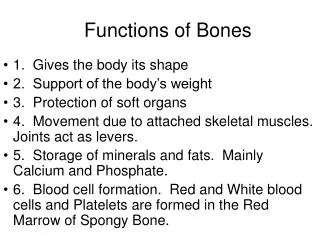
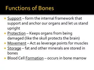
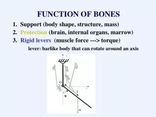
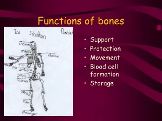
Function of Bones • form the framework that supports the body and cradles soft organs • provide a protective case for the brain, spinal cord, and vital organs • provide levers for muscles
Function of Bones • reservoir for minerals, especially calcium and phosphorus • hematopoiesis occurs within the marrow cavities of bones
Bone Markings • Bulges, depressions, and holes that serve as: • Joint surfaces • Conduits for blood vessels and nerves
Bone Markings: Projections – Sites of Muscle and Ligament Attachment • rounded projection • narrow, prominent ridge of bone • large, blunt, irregular surface • narrow ridge of bone • small rounded projection • raised area above a condyle • sharp, slender projection • any bony prominence
Bone Markings: Projections – Projections That Help to Form Joints • bony expansion carried on a narrow neck • smooth, nearly flat articular surface • rounded articular projection • arm-like bar of bone
Bone Markings: Depressions and Openings • canal-like passageway • cavity within a bone • shallow, basin-like depression • furrow • narrow, slit-like opening • round or oval opening through a bone
Bone Textures • Compact bone • Spongy bone • honeycomb of trabeculae _
Structure of Long Bone • Long bones consist of a _ • Diaphysis • Tubular shaft • Composed of _ • surrounds the medullary cavity • Yellow bone marrow in the medullary cavity
Structure of Long Bone • Epiphyses • ________________________________ of long bones • Exterior is compact bone, and the _ • Joint surface is covered with articular (hyaline) cartilage • Epiphyseal line separates the diaphysis from the epiphyses
Bone Membranes • ______________________________ – double-layered protective membrane • Richly supplied with nerve fibers, blood, and lymphatic vessels, which enter the bone via _ • Secured to underlying bone by _
Bone Membranes • delicate membrane covering internal surfaces of bone
Structure of Short, Irregular, and Flat Bones • Thin plates of periosteum-covered compact bone on the outside with endosteum-covered spongy bone on the inside • Have _ • Contain bone marrow between the trabeculae
Location of Hematopoietic Tissue (Red Marrow) • In infants • Found in the _ • all areas of spongy bone • In adults • Found in the _ • the head of the femur • the head of the _
Microscopic Structure of Bone: Compact Bone • _________________________, or osteon – the structural unit of compact bone • weight-bearing, column-like matrix tubes composed mainly of collagen • Haversian, or _ • containing blood vessels and nerves • channels lying at right angles to the central canal, connecting blood and nerve supply of the periosteum to that of the Haversian canal
Microscopic Structure of Bone: Compact Bone • Osteocytes • Lacunae • ______________________________ in bone that _ • Canaliculi • ___________________________________ that connect lacunae to each other and the central canal
Chemical Composition of Bone: Organic • Osteoblasts • Osteocytes • mature bone cells • Osteoclasts • large cells that resorb or _
Bone Development • Osteogenesis and ossification – the _________________________________, which leads to: • The formation of the bony skeleton in embryos • Bone growth until early adulthood • Bone thickness, _
Formation of the Bony Skeleton • Begins at ______________________ of embryo development • Intramembranous ossification • bone develops from a _ • Endochondral ossification • bone forms by _
Intramembranous Ossification • Formation of most of the _
Stages of IntramembranousOssification • An _____________________________ appears in the fibrous connective tissue membrane • Bone matrix is secreted within the fibrous membrane • Woven bone and periosteum form • Bone collar of _
Stages of Intramembranous Ossification Figure 6.7.1
Stages of Intramembranous Ossification Figure 6.7.2
Stages of Intramembranous Ossification Figure 6.7.3
Stages of Intramembranous Ossification Figure 6.7.4
Endochondral Ossification • Begins in the _ • Uses ____________________________” as models for bone construction • Requires breakdown of hyaline cartilage prior to ossification
Stages of Endochondral Ossification • Formation of bone collar • Cavitation of the hyaline cartilage • spongy bone formation • Formation of the medullary cavity; appearance of _ • Ossification of the epiphyses, with hyaline cartilage remaining only in the epiphyseal plates
Postnatal Bone Growth • Growth in length of long bones • Cells of the epiphyseal plate proximal to the resting cartilage form three functionally different zones:
Functional Zones in Long Bone Growth • Growth zone • ____________________________________, pushing the epiphysis away from the diaphysis • Transformation zone • older cells enlarge, the matrix becomes calcified, cartilage cells die, and the _ • Osteogenic zone • new _
Hormonal Regulation of Bone Growth During Youth • During infancy and childhood, epiphyseal plate activity is stimulated by _ • During puberty, _ • Initially promote adolescent growth spurts • Later induce epiphyseal ___________________________, ending longitudinal bone growth
Response to Mechanical Stress • Wolff’s law • a bone grows or remodels _ • Observations supporting Wolff’s law include • Long bones are thickest midway along the shaft (where bending stress is greatest) • Curved bones are thickest where they are most likely to buckle
Bone Fractures (Breaks) • Bone fractures are classified by: • The _____________________________ of the bone ends after fracture • The _______________________________ of the break • The ______________________________ of the bone to the long axis • Whether or not the bones ends penetrate the skin
Types of Bone Fractures • bone ends retain their normal position • bone ends are out of normal alignment
Types of Bone Fractures • bone is broken all the way through • bone is not broken all the way through • the fracture is ___________________________________ of the bone
Types of Bone Fractures • the fracture is __________________________________to the long axis of the bone • Compound (open) • bone ends do not penetrate the skin
Common Types of Fractures • Comminuted • bone fragments into __________________________ common in the elderly • ragged break when bone is _____________________________________ common sports injury • broken bone portion pressed inward; typical skull fracture
Common Types of Fractures • Compression • ___________________________________; common in porous bones • epiphysis separates from diaphysis along epiphyseal line; occurs where cartilage cells are dying • incomplete fracture where one side of the bone breaks and the other side bends; _
Stages in the Healing of a Bone Fracture • Torn blood vessels hemorrhage • A mass of clotted blood (_______________) forms at the fracture site • Site becomes swollen, painful, and inflamed Figure 6.13.1
Stages in the Healing of a Bone Fracture • Fibrocartilaginous _ • Granulation tissue (soft callus) forms a few days after the fracture • __________________________________ and phagocytic cells begin cleaning debris Figure 6.13.2
Stages in the Healing of a Bone Fracture • The fibrocartilaginous callus forms when: • ___________________________________ migrate to the fracture and begin reconstructing the bone • Fibroblasts secrete __________________________________ that connect broken bone ends • Osteoblasts begin forming spongy bone
Stages in the Healing of a Bone Fracture • Bony callus formation • New bone trabeculae appear in the fibrocartilaginous callus • Fibrocartilaginous callus _ • Bone callus _____________________________________, and continues until firm union is formed 2-3 months later Figure 6.13.3
Stages in the Healing of a Bone Fracture • Bone remodeling • Excess material on the bone shaft exterior and in the medullary canal is removed • ________________ is laid down to reconstruct shaft walls Figure 6.13.4