Download
1 / 85
850 likes | 1.03k Views
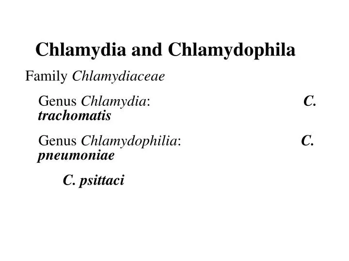
Chlamydia and Chlamydophila. Family Chlamydiaceae Genus Chlamydia : C. trachomatis Genus Chlamydophilia : C. pneumoniae C. psittaci. Chlamydiaceae. Obligate intracellular organisms Were once considered virus, true bacteria

E N D
Chlamydia and Chlamydophila Family Chlamydiaceae Genus Chlamydia: C. trachomatis Genus Chlamydophilia: C. pneumoniae C. psittaci
Chlamydiaceae Obligate intracellular organisms Were once considered virus, true bacteria Contain DNA and RNA Possess ribosomes, synthesize proteins, nucleic acid, and lipids, but cannot synthesize ATP. Binary fission Susceptible to numerous antibiotics, but not to penicillin (lack peptidoglycan) Cell wall: Major outer membrane protein (MOMP) – serological variants (serovars) Outer membrane protein 2 (OMP2) – cysteine-rich protein, structure stability of elementary body (EB)
Unique development cycle Two morphological distinct forms in cytoplasmic phagosome: (1) elementary body (300-400 nm), resistant to harsh environmental factors; bind to receptors of host cells and stimulate uptake; cannot replicate but infectious, (2) reticulate body (800-1000 nm), reproductive form, metabolically active, noninfectious. Histologic stains can detect phagosome with accumulated RBs (inclusion)
Life cycle of C. trachomatis Infectious Form Elementary Body (EB) Extrusion and release of EB Attachment & Ingestion Phagosome fusion Replication Form Reticulate Body (RB) Nucleus Condensation RB EB Multiplication of (RB)
1. Chlamydia trachomatis Infections only occur in humans Two biovarsand 18 serovars(antigenic differences in MOMP) BiovarsSerovars Disease Trachoma A to C Trachoma D to K Urethritis, cervicitis, Inclusion conjunctivitis, Neonatal conjunctivitis, Infant pneumonia LGV L1 to L3 Lymphogranulomavenereum (LGV)
Pathogenesis EBs enter the body via minute abrasions and lacerations Trachoma serovars primarily infect nonciliatedepithelial cells (urethra, endocervix, endometrium, fallopian tube, anorectum, respiratory tract, conjunctiva) LGV serovars replicate in mononuclear phagocytes (more invasive); formation of granuloma in lymph nodes draining the site of primary infection, abscesses, or sinus tracts formation
Pathogenesis Direct destruction of cells during replication Proinflammatory cytokine response stimulates a severe inflammation (accumulations of neutrophils, lymphocytes and plasma cells). No long-lasting immunity after infection Re-infection induces a vigorous inflammatory response with subsequent tissue damage (blindness and sterility).
Trachoma A chronic keratoconjunctivitis caused by serovars A, B, Ba, C. Diffuse follicular conjunctivitis → eyelid inward → eyelashes abrade cornea → corneal ulceration → pannus formation(invasion of vessels into the cornea) → blindness Endemic in the Middle East, North Africa, and southern Asia (crowded and poor sanitation regions); predominantly in children. Leading global causes of preventable blindness (>150 million infected, 6 million blinded). Transmission: eye-to-eye by droplet, hands, contaminated clothing, flies.
Urogenital infections Venereal infections caused by serovars of D to K. The most common sexually transmitted bacterial disease in U.S. 2.8 million new cases annually (50 million worldwide). In women: 80% asymptomatic as reservoir; bartholinitis, cervicitis, endometritis, salpingitis, urethritis, which can lead to sterility and ectopic pregnancy. In men: 25% asymptomatic; nongonococcalurethritis (NGU; urethritis caused by pathogens other than gonococcus )
NongonococcalUrethritis (NGU) C. trachomatis (35-50% of cases) Ureaplasmaurealyticum(10-30% of cases) Mycoplasmahominis Gardnerellavaginalis Trichomonasvaginalis Candida albicans
Dual infections of C. trachomatis and Neisseria gonorrhoeae are common. Post-gonococcus urethritis Symptoms of chlamydial infection develop after successful treatment of gonorrhea. Reason: longer incubation period + β-lactam antibiotics are ineffective for C. trachomatis Reiter’s syndrome Urethritis, conjunctivitis, polyarthritis, mucocutaneous lesion Usually occurs in young white man Initiated by genital infection with C. trachomatis.
Adult Inclusion Conjunctivitis Acute follicular conjunctivitis with mucopurulent discharge Mostly occur in sexually active adults (18-30 yr) with genital infection with serotypes A, B, Ba, D to K. Acquired by auto-inoculation, oral-genital contact
Newborn Inclusion Conjunctivitis 25% infants acquired from mothers with active genital infections Swollen and hyperemic eyelids Long (>12 months) disease course if untreated and are at risk for C. trachomatis pneumonia
Infant Pneumonia A diffuse interstitial pneumonia Occur in 10-20% infants that exposed to the pathogen at birth Rhinitis → staccato cough
Lymphogranulomavenereum (LGV) A chronic sexually transmitted disease caused by C. trachomatis L1, L2, L2a, L2b, L3. More common in men, with male homosexuals being the major reservoir. Small, painless lesions (papule or ulcer) at site of infection (genitalia). Fever, headache, myalgia. Inflammation and swelling of regional lymph nodes (inguinal nodes), painful buboes, rupture, fistulas formation. Proctitis is common in women. Resolve spontaneously or progress to ulceration or genital elephantiasis
Lab diagnosis Symptomatic infections are easier to diagnosis than asymptomatic infections as more chlamydiae present in specimen. Cytology – Giemsa-stained cell scrapings Quality of the specimen is important. Specimens must be obtained from the involved site; pus or exudate is inadequate. Insensitive, nonspecific Culture – HeLa, MaCoy, Hep-2 cells Iodine stain to detect inclusions (=RBs) The most specific methods for diagnosis. Sensitivity depends on quality and quantity of specimen.
Iodine-stained Chlamydia trachomatis inclusion bodies (arrows)
Lab diagnosis • Fluorescent antibody assay • Frei test (delayed hypersensitivity) for LGV • Growth in tissue culture • DNA probe test • PCR
C. trachomatisnew detection method PCR-SB Cryptic plasmid –PCR hybridization assay DNA purification PCR (DNA amplification) Hybridization (Oligo-probes)
DNA purification Phenol-CIA method
PCR (DNA amplification) Cryptic plasmid Two primers named CT-CPF: 5'‑TGATTGTACAAGGGATCCGTAAGT‑3' (start at nt. 7089) and CT-CPR: 5'‑TCGATGAAAGACAGGAAATACG‑3' (end at nt. 7465) (X07547, GenBank) Amplify 376 bps CPF CPR homology to some other genes of Human, Drosophila melanogaster andS.cerevisiae.
Hybridization (Oligo-probes) C. trachomatis-specific anti-sense probe named CP35 (nt.7335- nt. 7360) The oligoprobe, CP-35 has also sequence homology in many sequences of human.
Comparison of the results with PCR-microwell plate (MWP) assay and the PCR-SB assay using serially diluted positive sample -1 -2 -3 -4 -5 -6 Dilution of a positive sample 1 10 10 10 10 10 10 Amplicore MWP (OD) 3.03 2.68 0.70 0.51 0.30 0.07 0.08 PCR - SB 400 (bps) 300 (bps)
Comparison of the results with IDEIA, PCR-MWP and PCR-SB assays in 16 samples from urethritis patients Case No. 1 2 3 4 5 6 7 8 9 10 11 12 13 14 15 16 - - - - - - - - + + + + + + + + IDEIA Amplicore MWP (OD) 0.06 0.65 0.15 0.14 2.47 0.09 0.10 1.74 3.42 3.13 0.81 3.23 2.93 0.79 3.37 3.36 PCR - SB 400 (bps) 300 (bps) Agarose gel 400 (bps) 300 (bps)
T/P/C Doxycycline for LGV Azithromycin or doxycycline for ocular and genital infections in adult Erythromycin for newborn conjunctivitis and pneumonia Improve sanitary conditions – essential for prevention Safe sex practices
2. Chlamydophiliapneumoniae Was first isolated from the conjunctiva of a child in Taiwan - TWAR strain. An important cause of sinusitis, pharyngitis, bronchitis, and pneumonia. Infection is common, especially inadults and transmitted person-to-person by respiratory secretions.
Clinical disease Most infections are asymptomatic or mild - persistent cough. Cannot be differentiated with other atypical pneumonia - Mycoplasmapneumoniae, Legionellapneumophila, and respiratory viruses. Detected in atherosclerotic lesions in blood vessels. However, the role in the development of atherosclerosis is not clear.
Lab diagnosis Diagnosis is difficult Do not grow in cell lines used for isolation of C. trachomatis NAATs are OK with large interlaboratary variation. Micro-immunofluorescence (MIF) test The only acceptable serodiagnotic test (specific) A single IgM titer > 1:16 or a fourfold increase in IgG titer Nucleic Acid Amplification Techniques (NAAT)
T/P/C Ubiquitous present, control is difficult Macrolides (erythromycin), doxycycline
3. Chlamydophiliapsittaci Caused Psittacosis (parrot fever). The natural reservoir is any species of birds Can infect sheep, goat, cows, and humans (zoonosis) High risk groups: veterinarians, zookeepers, pet shop workers, employees of poultry industry.
Pathogenesis Inhalation of dried bird excrement, urine, or respiratory secretions; person-to-person transmission is rare. Bacteriafirstspread to and multiply in reticuloendothelial cells of liver and spleen necrosis Then hematogenous spread to lung and other organs via circulation Lmphocytic inflammation in lung, edema, necrosis, mucous plugs in bronchioles cyanosis and anoxia
Clinical disease Asymptomatic infection Flu-like illness: high fever, headache, chills, myalgia Serious pneumonia: non-productive cough, rales, consolidation, CNS involvement: common (headache, encephalitis, convulsion, coma) GI symptoms: nausea, vomiting, diarrhea (carditis, hepatomegaly, splenomegaly)
Diagnosis and treatment for C. psittaci Complement fixation test of paired acute and convalescent phase sera Confirmed by species-specific MIF test Treatment: tetracyclines or macrolides No need of isolation of patients and prophylaxia No vaccine available Treat birds with chlortetracycline HCl for 45 days.
Rickettsia and OrientiaEhrlichia, Anaplasma, Coxiella Rickettsia Howard Ricketts Ehrlichia Paul Ehrlich Coxiella Harold Cox (Historically classified in Rickettsiaceae)
Order Rickettsiales Family RickettsiaceaeGenera RickettsiaOrientia Family AnaplasmataceaeGenera EhrlichiaAnaplasmaNeorickettsiaWolbachia
Rickettsia and Orientia Obligate intracellularparasites. G(-) bacilli, with a minimal peptidoglycan layer (stain poorly with Gram stain) and LPS (weak endotoxin activity) Maintain in animal and arthropod reservoirs (by transovarian transmission). Transmitted to humans by arthropod vectors (ticks, mites, lice, fleas). Humans are accidental hosts: acquired by arthropod bite or contact of arthropod excreta with abraded skin
Rickettsia (also Ehrlichia) is unstable and die quickly outside host cells. Coxiellahighly resistant to desiccation, remain viable in environment for months to years. After phagocytosis Rickettsia and Orientia: degrade phagosome membrane by producing phospholipase, multiply in cytoplasm and nucleus of endothelial cells Ehrlichia and Anaplasma: multiply in cytoplasmic vacuoles (=phagosomes) of hematopoietic cells Coxiella: multiply in phagolysosome of monocytes and macrophages
Important Rickettsial Diseases Spotted fever group (17 species related to human diseases) R. rickettsii RMSF (>90%) R. akariRickettsialpox (100%) Typhus group R. prowazekii Epidemic typhus (40-80%) R. typhiMurine typhus (50%) O. tsutsugamushi Scrub typhus (<50%) (Parentheses: % of rash, The distribution of rickettsial diseases (restricted area or worldwide) is determined by the distribution of the arthropod hosts/vectors. Rocky Mountain spotted fever (RMSF)
Pathogenesis No toxins, no immunopathology OmpA mediated binding to endothelial cells Rickettsia replicate in endothelial cells, cause cell damage and blood leakage, vasculitis, skin rash, microthrombi, focal ischemia, hemorrhage. Hypovolemia, hypoproteinemia, reduced perfusion, organ failure.
Rocky mountain spotted fever Have a restricted geographic and seasonal distribution, corresponding to tick activity. R. rickettsii is maintained in hard ticks (wood tick and dog tick) by transovarian transmission. Transmitted to humans by tick bite (need >6h to establish infection). High fever, chills, headache, skin rash (>90%, extremities to trunk) Respiratory failure, encephalitis, renal failure.
Rocky mountain spotted fever Have a restricted geographic and seasonal distribution, corresponding to tick activity. R. rickettsii is maintained in hard ticks (wood tick and dog tick) by transovarian transmission. Transmitted to humans by tick bite (need >6h to establish infection). High fever, chills, headache, skin rash (>90%, extremities to trunk) Respiratory failure, encephalitis, renal failure.
Diagnosis is urgent, the prognosis depends on the duration of illness (identify key clinical signs – rash); fatality 10-25% if untreated Culture: buffy coat of blood or skin biopsy; tissue culture or embryonated eggs (danger) Microscopy: Giemsa stain; FA for biopsy tissue specimens (rapid and specific) Serology: microimmunofluorescence (MIF), detect antibodies against MOMP and LPS antigens; both specific and sensitive Nucleic acid-based tests: PCR + gene sequencing of a variety of genes The traditional Weil-Felix test: not recommended for use
Treatment /Prevention/Control: Appropriate therapy would result in good prognosis (e.g., doxycycline) No vaccine Prevent tick bites (can survive for as long as 4 years without feeding)
Rickettsialpox R. akari Infections are transmitted to humans from rodents reservoir by bite of infected mites (transovarian transmission) Cosmopolitan distribution (New York City) Clinical disease – biphasic Papule at site of bite, ulceration, eschar formation (differentiate with cutaneous anthrax) High fever, severe headache, chills, sweats, myalgias, photophobia, generalized rash (100%), complete healing 2-3 wks.
Epidemic (louse-borne) typhus R. prowazekii Humans are the primary reservoir with person-to-person transmission by human louse (the bacteria kill the lice 2 to 3 wk after infection; no transovarian transmission). Epidemics occur among people living in crowded, unsanitary condition - war, famine, or natural disaster. High fever, severe headache, myalgias, skin rash (20-80%), complete recovery >3 months
Brill-Zinsser Disease Bacteria may remain for years. A recrudescent, mild form of epidemic typhus arising years after the initial attack.
Diagnosis: MIF test T/P/C: Tetracyclines, Chloramphenicol Louse-control A formaldehyde-inactivated vaccine is available
Endemic (murine) typhus R. typhi transmits to man from rodent reservoir hosts by the bite of rat flea and cat flea. Endemic all over the world, primarily in warm, humid areas. Fever, severe headache, myalgias, chills, skin rash (50%) on chest and abdomen for 3 weeks.
Diagnosis: IFA test T/P/C: Tetracyclines Pest control No vaccine