Download
1 / 15
190 likes | 401 Views
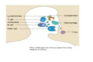
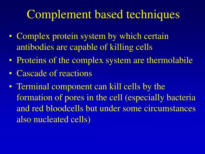
Complement based techniques. Complex protein system by which certain antibodies are capable of killing cells Proteins of the complex system are thermolabile Cascade of reactions

E N D
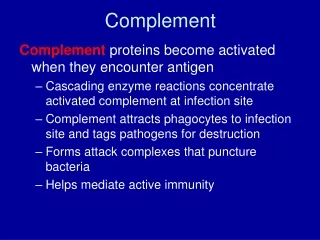
Complement based techniques • Complex protein system by which certain antibodies are capable of killing cells • Proteins of the complex system are thermolabile • Cascade of reactions • Terminal component can kill cells by the formation of pores in the cell (especially bacteria and red bloodcells but under some circumstances also nucleated cells)
Complement based techniques • Technique is applied in traditional serological HLA-typing • Antisera of persons who got immunized by circumstances against known HLA-antigens are used in a microcytotoxicity assay • Purify the cells to be HLA typed from the blood sample of a patient (T cells for HLA class I and B cells for HLA class II)
HLA-typing Fluorescein diacetate colours living cells green, propidium jodide colours dead cells red Analyse by microscope
HLA-typing with complement techn • Problems: • for some rare HLA-antigens no antiserum availale not detected • Cross-reactivity • In Class II typing: B-lymphocytes more sensitive to cross reactivity and non specific complement lysis • Less specific than analysis of MHC genes (LiPa strip, sequencing)
Line Immunoassay (LIA) • For the detection of antibodies in patients against other specific antigens (microorganisms or other antigens) (compare with latex agglutination) • The antigens are bound in strips on a nitrocellulose membrane • Detection of bound antibodies with enzym-labeled anti-human Ig (compare with ELISA, see later) substrate precipitation visible strip • Better specificity by simultaneous detection of several epitopes • Several commercial kits available: HIV, Hep C, Human T-cell Lymphotrope Virus (HTLV)
LIA BCIP/NBT (5-bromo-4-chloro-3-indoyl-phosphate / nitro blue tetrazolium) Antibody from serum of patient Antigen Alkaline phosphatase-labeled anti-human Ig
Western blotting • Used for detection of the presence of antibodies in for eg virology (compare with LIA) • The antigens are produced/purified and separated on a SDS-PAGE gel according to their molecular weight see figure • Can also be used for the detection of certain proteins (antigens) present in a sample • Separate total protein extract on a SDS-PAGE gel screen nitrocellulose membrane with specific (labeled) monoclonal antibodies or specific antiserum • eg. Test for the presence of prions in CJD and VCJD
Prion diagnosis in CJD and VCJD • Fast test (1 day) on a small amount of brain tissue • Proteinase K digest on SDS-PAGE nitrocellulose membrane screen with prion-specific Ab 3 bands of 16-31 kDa (non-mono-di-gly-PrPres) • Normal PrPc complete digestion by proteinase K
ELISA • Enzyme Linked Immunosorbent Assay • Can be used for the detection and quantitation of Ab’s • Can also be used for the detection and quantitation of antigen (proteins) • See figures • Very common technique many commercial kits are available
Flowcytometry • Very common technique in medical diagnosis especially in the field of hematology • Uses fluorescently labeled monoclonal antibodies to study the presence of certain specific markers on cells indication of disease sate or identification of certain cell types