Download
1 / 1
20 likes | 162 Views
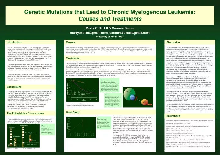
Chromosome 9 q+. 1.0. Chromosome 9. 0.9. 0.8. 0.7. Philadelphia Chromosome (or 22q-). Bcr-Abl. 0.6. Survival probability. 0.5. Chromosome 22. 0.4. Blocked. Total. Dead. 0.3. 261. 31. Imatinib mesylate. bcr-abl. ATP. 0.2. 251. 193. Others. bcr. 0.1.

E N D
Chromosome 9 q+ 1.0 Chromosome 9 0.9 0.8 0.7 Philadelphia Chromosome (or 22q-) Bcr-Abl 0.6 Survival probability 0.5 Chromosome 22 0.4 Blocked Total Dead 0.3 261 31 Imatinib mesylate bcr-abl ATP 0.2 251 193 Others bcr 0.1 (P<0.0001) FUSION PROTEIN WITH CONSTITUTIVE TYROSINE KINASE ACTIVITY P P 0 P P abl Imatinib mesylate 0 24 48 72 96 Months Fig. 3 Imatinib Mesylate binds to the Bcr-Abl kinase, blocking the ATP molecule from binding. Since the reaction between Bcr-Abl and ATP cannot occur, the cell never receives the signal to continue multiplying. This causes the apoptosy of the leukemic cell. Fig. 1 The Philadelphia Chromosome is a shortened chromosome 22 resulting from a translocation between chromosomes 9 and 22. Genetic Mutations that Lead to Chronic Myelogenous Leukemia: Causes and Treatments Marty O’Neill II & Carmen Banea martyoneillii@gmail.com, carmen.banea@gmail.com University of North Texas Introduction Causes Discussion Chronic Myelogenous Leukemia (CML) is defined as, “a malignant cancer of the bone marrow. It causes rapid growth of the blood-forming cells (known as myeloid precursors) in the bone marrow, peripheral blood, and body tissues.” [2] CML represents about 14% of all occurrences of leukemias. Patients who have CML are said to be in one of the following three phases (in order of occurrence): the chronic phase (between 1 and 15% blasts), the accelerated phase (between 15% - 30% blasts), and the blast phase (more than 30% blasts). [5] The chronic phase is the initial phase and the phase in which patients are most often diagnosed with CML [1]. The accelerated and blast phases are subsequent phases that represent further progression of CML in the patient and decreased chances of survival after treatment. Research concerning CML started in the XIX Century and is still in progress. There have been many important discoveries concerning CML regarding its causes, methods of detection in patients, treatments, and possible vaccines. Knowledge of Chronic Myelogenous Leukemia can be dated back to the mid-1800’s when symptoms of CML were first documented by Velpeau. Research continued, and about 20 years later, a high concentration of white blood cells in patients with CML was discovered. The term myelogenous was coined by Neuman after his 1878 discovery that bone marrow may not be the only place where blood cells were formed. In 1960, two scientists who lived in Philadelphia, Pennsylvania discovered what would later be called the Philadelphia (Ph) Chromosome: a direct indication of CML. The Philadelphia Chromosome results from a reciprocal translocation of parts of chromosomes 9 and 22 as is shown in Figure 1. This results in a shortened chromosome 22 called the Philadelphia Chromosome. Genetic mutations occur due to DNA damage caused by external agents such as ultraviolet light, nuclear radiation, or certain chemicals. [3] Mutations may also occur during the process of copying DNA in preparation for cell division. Even such common occurrences as exposure to benzene (produced by photocopy machines), in concentrations not greatly above the current legal standard [7], may ultimately lead to genetic mutations that trigger diseases such as CML. [6] Throughout our research we discovered various articles which linked potential carcinogenic substances (e.g. benzene) to the development of mutations in humans leading to various types of leukemias. Some of the articles dated back to 1981 [7], yet little action has taken place to adequately prevent human exposure to these harmful substances. In the case of benzene, the maximum legal exposure level is dangerously close to the concentration level which has been found to cause genetic mutations in humans. The patient in the case study was exposed to benzene while working in a copy center for 5 years. During this period it is likely that the patient suffered the mutation that ultimately triggered CML, since the diagnosis followed shortly thereafter. Despite the fact that employers are advised that employees might be exposed to carcinogenic substances while working in a certain environment, few of them respond appropriately. Therefore stricter regulations should be enforced by the government, in particular OSHA, to ensure that employees are adequately protected. Development of STI-571 opens the door to the further development of anticancer agents which are designed to target specific molecular abnormalities in human cancer. Before development of this drug, chemotherapy was a common option used by healthcare practitioners for treating CML. Chronic stage patients are no longer restricted to following the harsh treatment of chemotherapy including prolonged hospital stays, intravenous injections, and severe side effects. Despite progress in CML treatment, many of the patients experience resistance to STI-571 either from the beginning or throughout the course of their treatment. New research is needed to discover how to counteract the resistance. Several new drugs are currently in phase three clinical trials (conducted by the FDA) that may prove to be more effective for treating patients with resistance to STI-571. Throughout our research we found that interest was focused on the shortened 22nd chromosome abnormality, yet nothing was mentioned regarding the elongated 9th. It would be interesting to find out whether further mutations are induced by the elongated 9th chromosome, and what genes are potentially at risk. [1] D’Antonio, J. Chronic Myelogenous Leukemia. Clinical Journal of Oncology Nursing. 9(5): 535-8. [2] Faderl et al. Oncology (Huntingt). 1999; 13:169. [3] Genetic Science Learning Center at the University of Utah. http://gslc.genetics.utah.edu/. [4] National Marrow Donor Program overview slide presentation. http://www.marrow.org/NMDP/SLIDESET/sld031.htm. [5] Medline Plus Medical Encyclopedia. http://nlm.nih.gov. [6] Pasternak et al. Chronic Meylogenous Leukemia: Molecular and Cellular Aspects. J Cancer Res Clin Oncol. 1998; 643-60. [7] Rinsky et al. Leukemia in Benzene Workers. Am J Ind Med. 1981; 2(3): 217-45. [8] Smith, MT & Zhang, L. Biomarkers of Leukemia Risk: Benzene as a Model.Environ Health Perspect. 1998 Aug; 106 Suppl 4:937-46. [9] STI-571 in Chronic Myelogenous Leukaemia. British Journal of Haematology. 2002; 15-24. Treatments There are four principal therapeutic options which are mainly classified as: chemo therapy (hydroxyurea and busulfan), interferon, imatinib, and transplantation. While only transplantation usually leads to complete recovery, its drawbacks include a high risk of upfront mortality and limited availability depending on donor compatibility. [9] One of the newest discoveries in the field of cancer treatment is the development of STI-571 (Imatinib Mesylate), a milestone in modern pharmacology because of its ability to target specific molecules within cells (rational drug design). It was designed to bind to the Bcr-Abl tyrosine kinase molecule to impede its binding to the ATP (Adenosine 5’-triphosphate) molecule which would otherwise signal the leukemic cell to reproduce. This causes the leukemic cells to eventually die out by apoptosy. Fig. 2 Survival probabilities of patients in Chronic Phase CML, following α-Interferon failure. The probability of survival for those treated with Imatinib Mesylateis significantly higher than those who were not treated with it. Imatinib information is only available for the past 4 years (since its release). Background Accelerated and second chronic phase (n=744) National Marrow Donor Program overview slide presentation. [4] http://www.marrow.org/NMDP/SLIDESET/sld031.htm. Case Study The Philadelphia Chromosome References The patient was diagnosed with CML on December 21, 2004. The patient had a white blood count (WBC) performed in February, 2004, however, doctors did not conduct further tests despite anomalous WBC levels and distribution. Therefore, no additional tests were performed between February 2004 and December 2004. This is reflected in Figure 3 as a linear growth between these dates, despite the fact that it is likely that this growth would have been exponential due to normal cell division patterns. On December 15, 2004, the patient was found to have an enlarged spleen and had 4% blasts and 71,900 WBC, which indicated a potential diagnosis for CML. After bone marrow tests, the patient tested positive for BCR-ABL expression (resulting from Ph+-chromosome) which is a clear indication of CML. Fig. 4 White Blood Count progression in subject beginning one year before being diagnosed, and continuing throughout the first year of treatment with STI-571. [6] Pasternak et al. J Cancer Res Clin Oncol. 1998;124:643.