Download
1 / 19
190 likes | 611 Views
Chlamydia trachomatis , Mycoplasma , Ureaplasma , and other Non- Gonococcal urethritis:. Chlamydia trachomatis: Microscopy and culture: Small unicellular round-to-ovoid bacteria that cannot stained by Gram’s stain.

E N D
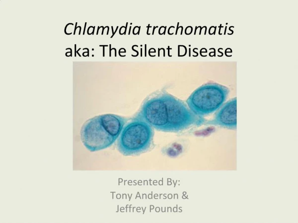
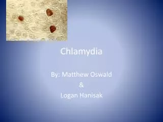
Chlamydia trachomatis, Mycoplasma, Ureaplasma, and other Non-Gonococcal urethritis: Chlamydia trachomatis: Microscopy and culture: Small unicellular round-to-ovoid bacteria that cannot stained by Gram’s stain. Someinclusion bodies retain Iodine or the counter stain safranin. Rigid Cell wall . The cell envelope has two lipid bilayers with cell wall material resembles a gram-negative (butnot peptidoglycan nor muramic acid).
n Obligatory intracellular parasite. It depends on the host cellular energy compounds ATP, and NAD. Cultivated in yolk sac of embryonated egg or tissue culture. Chlamydia inclusion :R. bodies. Chlamydia inclusion .
Pathogenesis and life cycle: -Transmission: Sexual route. -Infectiouspart: The elementary body. -The elementary bodies taken by phagocytosis into susceptible host cell. -Once inside the cell, the elementary body prevents fusion of the phagosome and lysosomes. -It will converted into metabolically active dividing Reticulate body. (non-infectious body). -Inclusion bodies. -After 48 hours, rupture of infected cell to release many elementary bodies. -Host cell death.
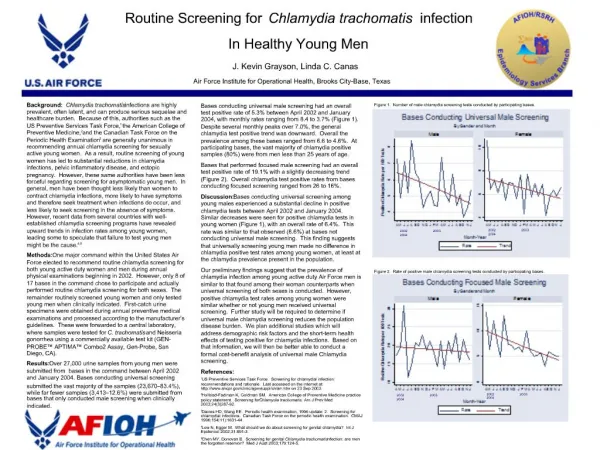
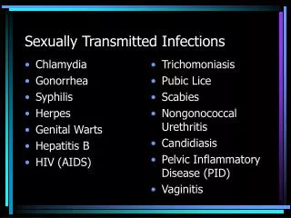
Clinical picture of Chlamydia trachomatis: Annually, more than four million urogenital Chlamydiatrachomatis infections occur in the USA in young individuals. 1-Nongonococcal urethritis: -Caused by Serovars: D,E, F,.., to K. - In male : Urethritis, infection could extend to epididymitis. and orchitis. - In Female: Pelvic inflammatory disease. Urethritis, Cervicitis, Endometritis, Salpingitis. 2-Lymphogranuloma venereum:(LGV): more invasive infection -Caused by Serovars: L1, L2, and L3. -Papules in the external genitalia.(for one to two months). -Painfulswelling of inguinal and perirectallymphnodes.
Clinical picture of Chlamydia trachomatis: Urethral discharge : (more mucoid with fewer pus cell). Chlamydial Cervicitis.
Laboratory diagnosis: Clinical specimens: Urethral discharge, urine, and Scraping of infected epithelial cells. 1- Direct microscopy: A-Immunofluorescent microscopy. B-Electron microscopy. 2-Detection of Chlamydia genetic material by PCR. 3-Serology: Serologic testing for specific antibodies is not helpful except in suspected Lymphogranulomavenereum.
Laboratory diagnosis: Immunofluorescent staining of inclusion body. Electron microscopy and immuno-electrone microscopy for inclusions.
Mycoplasma hominis and Ureaplasma urealyticum: The smallest prokaryotic microbe with no peptidoglycan cell wall. Because of their extremely small size(0.1-0.3 micrometer), Mycoplasma species pass through sterilization filters. Lacking cell walls, all species are enclosed instead by lipid bilayer membrane containing sterols. Due to the absence of Cell walls: 1-The bacteria are plastic, pleomorphic in nature, and cannot be classified as either coccior rods. 2-The bacteria are resistance to penicillin and cephalosporins.
n Double-stranded DNA genomes measure less than one million Kilodaltons. Cultural characteristics and colony morphology: Facultative anaerobes, and some species are strict anaerobes. Fastidious for external source of cholesterol (serum). Given appropriate supplementation, they can be grown in cell-free media. Colonies are visualized microscopically by 30 to 100 x magnification. Colonies show a characteristics (fried egg) appearance.
Biochemical activities and clinical picture: -Mycoplasma hominis and Ureaplasma urealyticum grow more rapidly than Mycoplasma pneumoniae. -They can be distinguished by their carbon utilization patterns; M. hominis degrades arginine. U. urealyticum hydrolyses urea. In female: -The major clinical condition associated with M. hominis is postabortal fever. -M. hominis is recovered locally in cases of Pelvic inflammatory disease. -All M. hominis species are Erythromycin resistance.
n The drug of choice for treatment is tetracycline (for M. hominis). Ureaplasmaurealyticum is associated with cases of Endometritis and vaginitis. In male: Ureaplasma urealyticum is associated with cases of Urethritis. The infection could be disseminated to other tissue in immunocompromised patients.
Candidiasis: - Most commonly encountered opportunistic mycoses worldwide. - Cellular immunity protects against mucocutaneous candidiasis, neutrophils protect against invasive candidiasis - They are members of the normal flora. - More than 150 species of Candida known. - Only ten species cause disease in humans. - The most common species of medical significance are: 1-Candidaalbicans. 2-Candidatropicalis.
Morphology and cultural characteristics: Candidais thin-walled, small yeasts (4 to 6 microns) that reproduce by budding. Microscopically: Candida albicansis dimorphic, in addition to budding yeast cells, pseudohyphae, it also can produce true hyphae. Asexual Germination of Candida occurs by production of Blastosporesor Chlamydiospores.
n -Macroscopically: on agar media they produce creamy colonies within 24 hours at 37 C or room temperature. -Candidaspecies produce a small ,white, rounded colonies with feet projection and regular margin. Germ tube test: -Candida species must be incubated with serum for 90 minutes at 37C; -yeast cells of C. albicanswill produce true hyphae or germ tube.
Clinical picture of Candida albicans: Candida albicanscauses almost 100% of cases of oropharyngeal candidiasisand at least 90% of cases of Candida vulvovaginitis. Vaginalcandidiasis presents as itching and burningpain of the vulva and vagina. Thick or thin white discharge. Vaginal swab and discharge should be examined for differential diagnosis. Candida albicans can cause urethritis in male.
Trichomoniasis: Trichomonas vaginalis: Classification: urogenital Mastigophora. Morphology: 20-30 um ,oval or pyriform in shape ,with short undulating membrane , axostyle and four free flagellae. Transmission:sexual intercourse,andcontaminated clothes. Pathology and Clinical picture: Vaginitis- itching, copious- yellowish offensive discharge. Urethritisin male and female. Prostatitisand seminal vesiculitis in male. Diagnosis: by finding the trophozoites in smears from vaginal or urethral discharge.