Download
1 / 32
320 likes | 510 Views
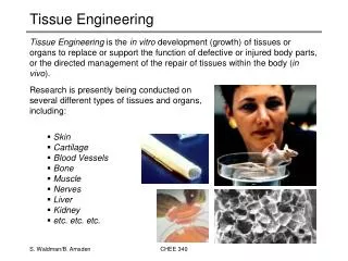
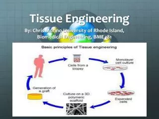
Techniques involved in tissue engineering. Mr. Viru Shah. Tuesday, August 3 rd , 2010. Preferred book . Content. Mammalian cell culture Cell identification Cell quantification Histology. Immunohistology Microscopy & SEM. 2. Mammalian cell culture.

E N D
Techniques involved in tissue engineering Mr. Viru Shah Tuesday, August 3rd , 2010
Content Mammalian cell cultureCell identificationCell quantificationHistology ImmunohistologyMicroscopy & SEM 2
Mammalian cell culture Important components in Cell culture Lab CO2 incubator Freezer (20-80 oC) Phase-contrast Microscopy Laminar Air Flow (BSL 2-3, depending on the sample) Cryotank 3
Mammalian cell culture Types of cells grown in culture Work area and equipment Cell Preservation and storage Cell Maintenance Safety considerations Tissue culture methods Asepsis is the base of any cell culture 5
Content Mammalian cell cultureCell identificationCell quantificationHistology ImmunohistologyMicroscopy & SEM 7
Cell identification Gross Morphological Molecular : RT- PCR OPN 146 PPARγ2 372 (Peroxisome proliferatoractivated receptor) (Osteopontin) Cellular : IF, FACS, Lentivirus IF: Immunofluorescence FACS: fluorescence activated cell sorter 8
Content Mammalian cell cultureCell identificationCell quantificationHistology ImmunohistologyMicroscopy & SEM 9
Cell quantification The yellow tetrazolium MTT (3-(4, 5-dimethylthiazolyl-2)-2, 5- diphenyltetrazolium bromide) is reduced by metabolically active cells, toan insoluble purple formazan crystals. This is solubilized and quantifiedby spectrophotometer Formazan crystal Absorbance of the converted dye is measured at a wavelengthof 570nm with background subtraction at 650nm. 10
Cell quantification Determination of cell numbers using a hemocytometer. Cells are countedafter staining with Trypan Blue. Dead cells stain, living cells do not. Hemocytometer Calculation: Average cell count per quadrant x dilution factor x 104 = Cells/ml 11
Content Mammalian cell cultureCell identificationCell quantificationHistology ImmunohistologyMicroscopy & SEM 12
Histology Histology is the study of tissue structure .To examine the tissue components. • Fixation • Sectioning • Visualization 13
Histology Fixation : Prevents its decomposition; cell metabolism is stopped. It is usually,but not always, done before sectioning The most common fixative is Formalin, a 37% aqueous solution offormaldehyde, which is either used on its own or in combination withother chemicals Formaldehyde reacts with the amino groups of proteins, and thuspreserves the general structure of the cell and extracellularcomponents 14
Histology Sectioning : To allow a specimen to be examined, it must be thinly sliced on thelevel of micrometers (1/1000th of a millimeter). Sectioning requires a microtome. The tissue is fixed, dehydrated andcompletely permeated with paraffin. It is then embedded in a paraffinblock. This block is stuck to a chuck, and then placed in a holder, whichis progressed by a rotating wheel. 15
Histology Visualization : When observing a freshly fixed section of tissue through a microscope,very little contrast is observed between adjacent regions of tissuewhich may have vastly different composition and function. As a result,individual cells or parts of cells are difficult or impossible to visualize. 16
Mammalian cell cultureCell identificationCell quantificationHistology ImmunohistologyMicroscopy & SEM 17
Immunocytochemistry Immunocytochemistry is technique to detect localized protein ormolecule by using antibodies Immunocytochemistry Screening for antibody reactivity New investigations Diagnosis and treatment Immunohistology = Immunochemistry of proteins in situ Immunohistochemistry = tissues = cells in their normal environment Immunocytochemistry = cells = cells isolated from tissues, cells in culture 18
Immunocytochemistry Peroxidase method: In this method the enzyme most often coupled to the anti-IgG isperoxidase, which is reacted with 3’ , 3’ diaminobenzidine (DAB) in thepresence of H2O2. This reaction gives an electron-dense deposit that canbe detected with light microscope. 19
Immunocytochemistry Fix cell & Permeabilize Expose to primary antibodies Secondary antibodies carrying tag 20
Immunocytochemistry UV illumination - immunofluorescence 21
Immunocytochemistry Immunocytology vs immunohistology Cells (better resolution by cytology) versus tissues(better expression context in tissues) Resolution of cellular component 23
Immunohistology Common problems in immunohistology Antibodies do not work as describes - Use reputable sources, complain to supplier, test them yourself Antibodies do not react with formalin-fixed section -Try frozen section, different fixatives, antigen retrieval methods;stronger antibody? polyclonal antibody High background on mouse tissues -Try blocking strategies, direct labeling 25
Mammalian cell cultureCell identificationCell quantificationHistology ImmunohistologyMicroscopy & SEM 26
Microscopy & SEM The observation of biological structure is made difficult by the factthat cell are in general, very small and transparent to visible light andthus are not visible to the unaided eye. Light microscopy Fluorescence microscopy Scanning electron microscopy 27
Microscopy & SEM In light microscopy resolving power depends upon the wave length ( λ )and the numerical aperture ( NA ) of objective lens. The limit of resolution, defined as the minimum distance between twopoints that allows for their discrimination as two separate point Limit of resolution = 0.61 λ NA With white light, the resolving power is about 250 nm (0.25 µm) The higher the resolving power ,the smaller the limit of resolution 28
Microscopy & SEM Light Microscope Bright field of human ileum 29
Microscopy & SEM 1. Energy is absorbed by the atom which becomes excited. 2. The electron jumps to a higher energy level. 3. Soon, the electron drops back to the ground state, emitting a photon (or apacket of light) - the atom is fluorescing. Immunofluorescence staining of epithelium cell (Hep-2) mitochondria Fluorescence microscope 30
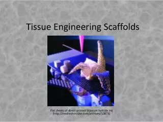
Microscopy & SEM Scanning electron microscopy (SEM) helps in studying the surface viewof the specimen employing secondary electron emission that is ejectedafter the primary electron beam has interacted with the surface of a thickspecimen Scanning electron microscope (SEM) Polymer Scaffold 31