Download
1 / 20
521 likes | 1.62k Views
Immunohistochemistry. Immunohistochemistry. ESE3. What is Immunohistochemistry?. a method used to detect and achieve visualization of the presence and distribution of a specific antigen in cells or tissues

E N D
Immunohistochemistry Immunohistochemistry ESE3
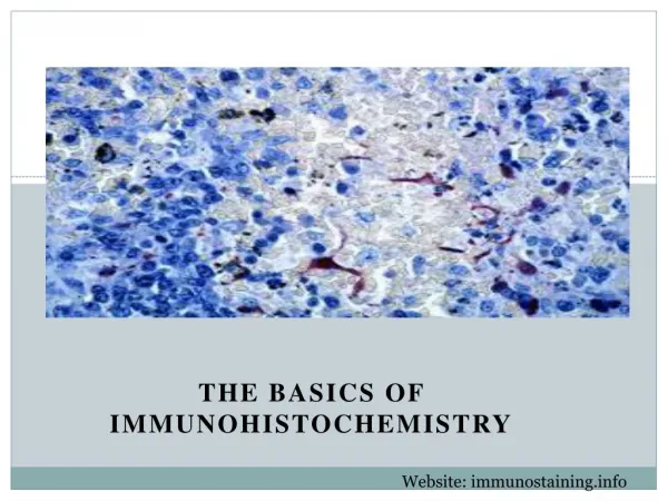
What is Immunohistochemistry? • a method used to detect and achieve visualization of the presence and distribution of a specific antigen in cells or tissues • uses antigen-antibody reactions, a ABC reagent and an enzyme substrate to achieve staining What I Did • stained prostate tissue samples for ESE3 • troubleshooted ESE3 antibody • using the controls • - no antibodies • - primary antibody only • - secondary antibody only • - IgG and secondary antibody
ESE3 • a nuclear protein • a transcriptional factor • epithelial-specific expression • expressed in normal cells • down-regulated in cancerous cells at both the mRNA and protein levels • acts as a tumor suppressor due to it’s involvement in the control of cell differentiation
Immunohistochemistry Protocol Immunohistochemistry Protocol
Part 1 Deparaffinize and Rehydrate 1. 3 x10 min xylene; 2 x 2 min in 100% ETOH, 2 x 2 min in 70% ETOH. Make sure to shake off excess xylene on slide before putting the slides into EtOH in order not to dilute the EtOH. 2. Rinse sides gently with ddH2O squirt bottle being careful not to rinse directly on the section. Wash with ddH2O in Coplin jar for 2 x 2 min on shaker at 120 rpm. Quenching Endogenous Peroxidase 1. Wipe off excess ddH2O from slide - work quickly to ensure that section does not completely dry out. To do this shake the slide and use a kimwipe to get rid of any extra liquid. 2. Pipette enough hydrogen peroxide (3% H2O2 in H2O) to cover the entire section and incubate for 10 min. Does not need to go into a humidity chamber. 3. Wash Sections with 1 x TBST with squirt bottle and then wash in Coplin jar for 2 x 2 min on shaker.
Antigen Retrieval • 1. While section is incubating in hydrogen peroxide, pre-heat Antigen • Retrieval Buffer in food steamer for 15 mins. • Lift up the containers in order to fill the bottom of the steamer up with tap water, up to the second line. • Put approximately 200 mL of antigen retrieval buffer into the glass slide holder in the steamer. • 2. Incubate slides in pre-heated Antigen Retrieval Buffer for 20 min. • Use forceps to transfer the slides into the slide holder. • 3. Remove lid from the food steamer after the 20 min period and let • the slides sit inside the steamer for 10 min. • 4. Remove the entire slide box with buffer and let sit at room • temperature on bench for 20 mins. • 5. Wash slides in 1 x TBST for 3 x 2 min in Coplin jar on shaker
Blocking and Primary Antibody Addition • 1. Gently dry slides and put in humidity chamber which is • tupperware lined with moist paper towels and a slide holder. • Redraw hydrophobic barriers, if they come off. • 2. Incubate slides for 1 hr at room temperature with blocking • buffer in the humidity chamber: 10% rabbit serum (RS) and • 1 x TBST. • 5 slides • 2x[1.00 mL of 1xTBST and 100 uL of RS] • 3 or 4 slides • 1.25 mL of 1xTBST and 125 uL of RS • 3. Wipe and shake off excess blocking buffer. Add primary antibody • diluted in blocking buffer and incubate overnight at 4 C in • humidity chamber • 5 slides (1:300) Rat IgG (1mg/mL) - 1:300 • 2x[3.33 uL of ESE3 in blocking buffer] 1.33 uL of Rat IgG in blocking buffer • 3 or 4 slides (1:300) • 4.16 uL of ESE3 in blocking buffer
Part 2 - Visualization with Vector Labs ABC Kit • 1. Wash slides 3 x 5 min in 1 x TBST • 2. Add appropriate biotinylated secondary antibody to each section. • Incubate in humidity chamber for 1 hr at room temperature. • For anti- rat biotinylated antibody (1:200) • 5 slides • 5 uL in 1.0 mL of 1x TBST • 3 or 4 slides • 6.25 uL in 1.0 mL of TBST • 3. During this incubation, prepare ABC reagent following • manufacturer’s instructions or for a small volume add 12.5 uL of • Reagent A to 1.25 mL of dilution buffer, TBST, then immediately add • 12.5 uL of Reagent B and mix well. Allow for ABC reagent to sit at • room temperature for about 30 min. • 4. Wash Slides for 2 x 5 min in 1 x TBST on shaker. • 5. Incubate sections with ABC reagent for 1 hr in humidity chamber. • 6. Wash slides for 2 x 5 min in 1 x TBST on shaker.
7. Prepare DAB solution and add appropriate amount to slides. • Incubate slides in DAB solution for 4 min • In 5.0 mL of distilled H2O • i. Add 2 drops of buffer, mix well • ii. Add 4 drops of DAB stock solution, mix well • iii. Add 2 drops of Hydrogen Peroxide, mix well • 8. Wash slides under running distilled H2O for about 30 sec. • Incubate in distilled H2O for 5 min. • 9. Counterstain with appropriate amount of haematoxylin [HE] for • 40 sec. Wash slides under running tap H2O for about • 30 sec. Incubate in tap H2O for 5 min. • 10. Clear and dehydrate slides: 2 x 3 min 70% EtOH, 2 x 3 min • 100% EtOH, 2 x 3 min xylene. • 11. Coverslip with Cytoseal mounting medium.
Results Results
Normal Cancerous PIN 40x 60x
Normal Gland (40x) nuclear staining
Cancerous Gland (40x) no nuclear staining
PIN (40x) no nuclear staining nuclear staining
Special Thanks To Dr. Tang & Judy Yan supervisor
Rick Hart - Principal of Hill Park Secondary School Mr. Watts Dr. Tang Dr. Tang's Graduate Students