Download
1 / 30
320 likes | 466 Views
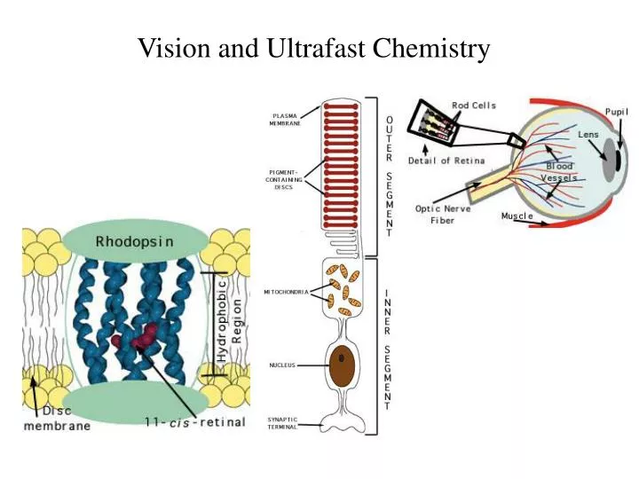
Vision and Ultrafast Chemistry. Visual signaling. Light. Rod. Cone. G-protein signaling pathway. Rhodopsin. Visual Receptor Protein Rhodopsin. Humphrey et. al., J. Molec. Graphics, 14 :33-38, 1996. Freely available, with source code from http://www.ks.uiuc.edu/Research/vmd/. Rhodopsin.

E N D
Visual signaling Light Rod Cone G-protein signaling pathway Rhodopsin
Visual Receptor Protein Rhodopsin Humphrey et. al., J. Molec. Graphics, 14:33-38, 1996 Freely available, with source code from http://www.ks.uiuc.edu/Research/vmd/
Rhodopsin Bacteriorhodopsin GPCR, vision in all species Photosynthesis, proton pump
Organization of the Purple Membrane of Halobacteria hn H+ V Baudry et al, J. Phys. Chem. (in press) assembly Ben-Nun et al Faraday Disc. 110: 447-462 (1998) Molnar et al J. Mol. Struct. (in press) function protein molecular electronics
Constructing and Simulating the Purple Membrane assembly function protein • Vibrational Spectroscopy (Kyoto) • Organic Synthesis (Rehovot) • Quantum Chemistry (Heidelberg) • Photophysics (Siena) • Protein Simulation (Urbana) • Pharmacolgy (New York) molecular electronics
Molecular Dynamics Program Used: NAMD2 hexagonal unit cell 23700 atoms per unit cell # processors Periodic boundary conditions in 3D (multilayers); NpT (constant pressure) simulations; Particle Mesh Ewald (no electrostatic cutoff); ~2 weeks/ns on 4 Alpha AXP21264-500Mhz procs.
Thermodynamics of the Purple Membrane PM thickness NpT simulation: constant temperature, variable volume In-plane dimensions Reduction of PM thickness during NpT simulation
Distribution of external water after MD Asp96 retinal Arg82 Equilibration of PM: rearrangement of water molecules Before MD After MD water Nb of atoms protein “c” dimension perpendicular to the membrane Top view of PM: Water molecules penetrate the PM, but not the protein, stop at Arg82 & Asp96
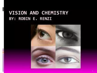
Color in Vision cone cells Visual receptors of rhodopsin family are classified based on their color sensitivity
Rhodopsin Family of Proteins • Seven transmembrane helices • Retinal chromophore bound • to a lysine via the Schiff base protonated Schiff base retinal (PSBR)
Color Regulation Visual receptors detect light by electronic excitation of retinal at different wavelengths. 400nm 500nm 600nm Absorption spectra of retinal in different visual receptors Question: How does the protein tune the absorption spectrum of retinal?
Spectral Tuning in Archaeal Rhodopsins sRII hR bR sRI 500nm 600nm Sensory Rhodopsin II (sRII) Repellent response to blue-green light Spectral features • Absorption maximum • is strongly blue-shifted • (70 nm from bR). • Prominent sub-band.
X-ray Structures of bR and sRII Landau et al. Unique opportunity to study spectral shift given by the availability of X-ray structures. • Structures are homologous. • (e.g., all-trans retinals) • Spectra are significantly • different. orange: sRII (Natronobacterium pharanois) purple: bR (Halobacterium salinarum)
Binding Sites of bR and sRII • Similar structure • Aromatic residues. • Hydrogen-bond network. • (counter-ion asparatates, • internal water molecules) bR Mutagenic substitutions (Shimono et al.) sRII T204A/V108M/G130S of sRII produces only 20 nm (30%) spectral shift. What is the main determinant(s) of spectral tuning?
Calculation of Absorption Spectra of bR and sRII • Combined quantum mechanical/molecular • mechanical (QM/MM) calculations. • Retinal is described by • ab initio MO (HF/CASSCF). • Protein environment by • molecular mechanics force • field (AMBER94).
Mechanism of Spectral Tuning S0 S1 S2 • Electrostatic interaction between • the retinal Schiff base and protein S2 positive charge S2 S1 S1 + + S0 O O C Asp (Glu) S0 isolated in protein • Electronic reorganization of retinal • due to polarization of retinal’s wave function
Results sRII bR 500nm 600nm S1-S0: 6.1 (exp. 7.2) kcal/mol. (shift of main absorption band) The shift is mainly due to electronic reorganization. S2-S1: 1.7 (exp. 4.0) kcal/mol. (appearance of side band in sRII) Optically forbidden in bR, but a peak (side-band) appears in sRII due to intensity borrowing from the S1 state, which is optically allowed.
Structural Determinants of Spectral Shift G helix is displaced in sRII. Distance between the Schiff base and the counter-ion is shorter. G helix N16 – Cg (Asp201: sRII) : 4.5 A N16 – Cg (Asp212: bR) : 5.2 A QM/MM optimized structures orange: sRII, purple: bR
Rhodopsin Photodynamics Quantum (Wave Packets) Dynamic in protein, 1-dimensional surface Ben-Nun et al., Faraday Discussion, 110, 447 - 462 (1998)
On-the-flyab initioQM/MMMD Simulation • An analogue of retinal (three • double bonds) in bR (20 QM • atoms, 96 basis functions) • CASSCF (6,6) / AMBER
The Role of Conical Intersection Topographyon the Photoisomerization of Retinal Michal Ben-nun Emad Tajkhorshid Shigehito Hayashi Jerome Baudry $$: Beckman Institute, NSF, HFSP, NIH-NCRR