Download
1 / 65
710 likes | 917 Views
Bones of the Upper Limb. Dr. Fadel Naim Orthopedic Surgeon Faculty of Medicine IUG-Gaza. لَا أُقْسِمُ بِيَوْمِ الْقِيَامَةِ{1} وَلَا أُقْسِمُ بِالنَّفْسِ اللَّوَّامَةِ{2} أَيَحْسَبُ الْإِنسَانُ أَلَّن نَجْمَعَ عِظَامَهُ{3} بَلَى قَادِرِينَ عَلَى أَن نُّسَوِّيَ بَنَانَهُ{4}.

E N D
Bones of the Upper Limb Dr. Fadel Naim Orthopedic Surgeon Faculty of Medicine IUG-Gaza
لَا أُقْسِمُ بِيَوْمِ الْقِيَامَةِ{1} وَلَا أُقْسِمُ بِالنَّفْسِ اللَّوَّامَةِ{2} أَيَحْسَبُ الْإِنسَانُ أَلَّن نَجْمَعَ عِظَامَهُ{3} بَلَى قَادِرِينَ عَلَى أَن نُّسَوِّيَ بَنَانَهُ{4}
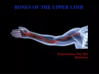
Skeleton of the Upper Limb • Each upper limb has 32 bones • Two separate regions • 1. The pectoral (shoulder) girdle (2 bones) • 2. The free part (30 bones)
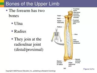
Upper Limb • The pectoral girdle consists of two bones: • the scapula • the clavicle • The free parthas 30 bones • 1 humerus (arm) • 1 ulna (forearm) • 1 radius (forearm) • 8 carpals (wrist) • 19 metacarpal and phalanges (hand)
Pectoral Girdle clavicle scapula humerus
Clavicle • The clavicle is an s-shaped bone that attaches the trunk to the upper extremity • Medial 2/3 convex forward and lateral 1/3 concave forward
Clinical Connection – Fractured Clavicle • A fall on an outstretched arm (F.O.O.S.H.) injury can lead to a fractured clavicle • The clavicle is weakest at the junction of the two curves • Forces are generated through the upper limb to the trunk during a fall • Therefore, most breaks occur approximately in the middle of the clavicle
Acromial end • is flat and has a small facet for articulation with the acromion • Sternal end • has a large facet for articulation with the manubrium, and first costal cartilage • Conoid tubercle • Attachment of conoid ligament of the coracoclavicular ligament • Trapezoid line • Attachment of trapezoid portion of the coracoclavicular ligament
Clavicle: Ligament Attachments • Sternal end of clavicle to first costal cartilage: Costoclavicular ligament • Conoid tubercle: Conoid portion of coracoclavicular ligament • Trapezoid line: Trapezoid portion of coracoclavicular ligament
Clavicle: Muscle Attachments • Deltoid • Pectoralis major • Trapezius • Sternocleidomastoid • Subclavius
Muscular, ligamentous, and fascial attachments to the clavicle
Scapula • Flat triangular bone • On the posterior thoracic wall • Between 2nd and 7th rib
AnteriorScapula • Borders: • Superior • Medial • lateral • Angles: • Superior • inferior • coracoid process • acromion • neck of scapula
Anterior Scapula acromion process coracoid process glenoid cavity superior angle subscapular fossa inferior angle
PosteriorScapula • Spine of scapula • Divides the supraspinous and infraspinous fossae • Serves as attachment for the deltoid and trapezius • Acromion: • Lateral extension of spine of scapula; • Articulate with clavicle • Greater scapular notch • Point at which the spine of the scapula ends, but the acromion continues; • Coracoid process • Partially seen as it projects anteriorly;
Supraspinous fossa • Origin of the supraspinatus muscle • Infraspinous fossa • Origin of the infraspinatous muscle • Lateral border • Attachment of: • Teres major • The long head of the triceps brachii • Teres minor
Posterior Scapula acromion process supraspinous fossa infraspinous fossa spine lateral border medial border
LateralScapula • Supraglenoid tubercle • Attachement of the long head of the biceps brachii • Infraglenoid tubercle • Attachement of the long head of the triceps brachii
LateralScapula • Acromion: • Articulates with the clavicle • Attachment for the trapezius and deltoid muscles; • Superior and inferior angles • Coracoid process: • Attachment point for: • The short head of the biceps brachii • Corachobrachialis • Pectoralis minor
The Humerus • Longest and largest bone of the free part of the upper limb • The proximal ball-shaped end articulates with the glenoid cavity of the scapula • The distal end articulates at the elbow with the radius and ulna
The Humerus • The proximal end consists of: • The head • Anatomical neck • Greater and lesser tubercles separated from each other by an intertubercular groove (bicipital groove)
The head • The head, nearly hemispherical in form • Articulates with the glenoid cavity of the scapula. • The circumference of its articular surface is slightly constricted and is termed the anatomical neck
The anatomical neck • The anatomical neck of the humerus is an indentation distal to the head of the humerus on which the articular capsule attaches. • Fracture of the anatomical neck rarely occurs.
The Greater Tubercle • The greater tubercle is situated lateral to the head and lesser tubercle, and just lateral to the anatomical neck • Its upper surface is rounded and marked by three flat impressions: • the highest for insertion of the suprasinatus muscle • the middle for the infraspinatus muscle • the lowest one, and the body of the bone for teres muscle
The Lesser Tubercle • The lesser tubercle is more prominent than the greater tubercle • Above and in front it presents an impression for the insertion of the tendon of the subscapularis muscle
The Intertubercular (Bicipital) Groove • The tubercles are separated from each other by a deep groove, the intertubercular groove (bicipital groove), which lodges the long tendon of the biceps brachii muscle
The surgical neck • The surgical neck is the point distal to the tubercles at which the superior portion of the bone meets the shaft • The surgical neck is a common site of fracture.
The body of the humerus has two prominent features: • The deltoid tuberosity, laterally, for attachment of the deltoid muscle
Distal Humerus • Medial epicondyle: • The pronator and flexor muscles of the forearm originate here • Lateral epicondyle: • The extensor and supinator muscles of the forearm originate here • Medial supracondylar ridge • Lateral supracondylar ridge • Trochlea (medial condyle): • Articulates with the trochlear notch of the ulna • Capitulum (lateral condyle): • Articulates with the radial head
Distal Humerus • Coronoid fossa: • Accommodates the coronoid process of the ulna during flexion. • A fat pad is situated here • Radial fossa: • Accommodates the head of the radius during flexion. • A fat pad is situated here • Olecranon fossa: • Accommodates the olecranon. • A fat pad is situated here • Groove for ulnar nerve
Anterior Humerus lesser tubercle medial epicondyle deltoid tuberosity trochlea coronoid fossa intertubercular groove greater tubercle capitulum lateral epicondyle
Humerus: Anterior • Clavicle • Acromion process • Greater tubercle • Coracoid process • Lesser tubercle • Scapula • Glenoid fossa • Humerus • Coronoid fossa • Deltoid tuberosity • Medial epicondyle • Lateral epicondyle • Capitulum • Trochlea • Radius • Ulna • Click R Button for Slideshow
Humerus: Anterior2 • Clavicle • Acromion process • Greater tubercle • Coracoid process • Lesser tubercle • Scapula • Glenoid fossa • Humerus • Coronoid fossa • Deltoid tuberosity • Medial epicondyle • Lateral epicondyle • Capitulum • Trochlea • Radius • Ulna
Humerus: Posterior • Acromion of scapula • Head of humerus • Spine of scapula • Greater tubercle • of humerus • Scapula • Glenoid fossa of scapula • Deltoid tuberosity • of humerus • Humerus • Medial epicondyle • of humerus • Lateral epicondyle • of humerus • Olecranon process of ulna • Radius • Ulna • Right Arm, Posterior
Radius • The radius is the lateral and shorter of the two forearm bones. • Its proximal end consists of: • A short cylindrical (or thick disc like) head • The smooth superior aspect of the head of the radius is concave for articnlation with the capitulum of the hnmerus during flexion and extension of the elbow joint. • The head also articulates peripherally with the radial notch of the ulna • The head is covered with articular cartilage. • A neck • Relatively constricted between the head and the tuberosity. • A medially directed tuberosity • The oval radial tuberosity separates the proximal end of the radius from the body.
The distal end of the radius • Its medial aspect forms a concavity, the ulnar notch, which accommodates the head of the ulna. • Extending from its lateral aspect is the radial styloid process. • The dorsal tubercle, (Lister’s) projecting dorsally lies between grooves for the passage of the tendons of forearm muscles
The radial styloid process is much larger than the ulnar styloid process and extends approximately a finger's breadth further distally • This relationship is of clinical importance when the ulna and/or the radius are fractured
Radius radial tuberosity head styloid process
Ulna • The stabilizing bone of the forearm is the medial and longer of the two forearm bones • Its proximal end has two prominent projections: • The olecranon: • projects proximally from its posterior aspect • The coronoid process • Projects anteriorly.
The olecranon • The olecranon is the most proximal posterior eminence of the ulna • It is on the dorsal subcutaneous border and contains broad attachments for the triceps posteriorly • Anteriorly, the olecranon forms the trochlear notch of the ulna, which articulates with the trochlea • The radial notch • On the lateral side of the coronoid process is a smooth, rounded concavity, which articulates with the head of the radius.
Ulna • Inferior to the coronoid process is the tuberosity of the ulna for attachment of the tendon of the Brachialis muscle. • Inferior to the radial notch on the lateral surface of the ulna is a prominent ridge (the supinator crest) • Between it and the distal part of the coronoid process is a concavity (the supinator fossa) • The deep part of the supinator muscle attaches to the supinator crest and fossa.
styloid process • At the narrow distal end of the ulna is a abrupt enlargement forming a disclike head and a small, conical styloid process.