Download
1 / 34
380 likes | 607 Views
The Heart. YC Wong, PhD 王雲川 The University of Hong Kong ycwong@hku.hk. Scope. Overview of anatomy of the heart Surface markings and gross anatomical structure Vascular system of the heart Impulse conducting system Histology of the heart. Location and surface markings.

E N D
The Heart YC Wong, PhD 王雲川 The University of Hong Kong ycwong@hku.hk
Scope • Overview of anatomy of the heart • Surface markings and gross anatomical structure • Vascular system of the heart • Impulse conducting system • Histology of the heart
Location and surface markings • Located in middle mediastinum 纵隔 • Locate the sternal angle • Point one: right 3rd costal cartilage 1 cm from sternum border • Point 2: 6th costal cartilage, 1 cm from sternum • Point 3: 5th left intercostal space just medial to mid clavicular line • Point 4: 2nd left intercostal space at the border of sternum • Join the 4 points to mark the outer shape of the heart Sternal angle
The Pericardium 心包膜 • Fibrous capsule enclosing the heart • Separated by pericardial cavity filled with fluid • To reduce the friction of heart during pumping • Lined by serous pericardium 浆膜心包 • Subdivided into parietal and visceral layers • The parietal layer closely adheres to fibrous pericardium 纤维心包; the visceral layer reflected on the outer surface of heart, the external limit of epicardium 心外膜, lined by mesothelium 间皮 • Perforated only at the roots of major vessels • Reflections of serous pericardium forms transverse sinus 横窦and oblique sinus 斜窦
The Pericardium 心包膜 Trans Sinus L pulmonary V R. Pulmonary V Oblique Sinus
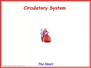
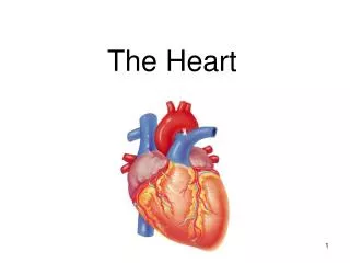
Anatomy of the Heart • Located in middle mediastinum • Surrounded by pericardium • Apart from attachment to major vessels, the rest is basically free within pericardial cavity • Three surfaces: sternocostal surface, diaphramatic surface and base, i.e. the posterior surface • The apex心尖 of the heart is directed forward, downward and to the left Left auricle Right atrium Left ventricle Right ventricle Apex
Four Chambers • Divided by vertical septa to form 4 chambers • Right atrium and left atrium, separated by interatrial septum • Right ventricle and left ventricle, separated by interventricular septum • Right atrium and right ventricle is guarded by a valve, the tricuspid valve, 三尖瓣 • Left atrium and left ventricle is guarded by mitral valve 二尖瓣 • Deoxygenated blood returns to right atrium then to right ventricle which is pumped to the lungs via pulmonary trunk, guarded by pulmonary valve 肺动脉瓣 • Oxygenated blood from the lungs enters the left atrium, then to left ventricle • This blood is pumped through aorta guarded by aortic valve 主动脉瓣 Right Atrium Left atrium Pulmonary v Aortic v Mitral v Tricuspid v Left ventricle Right ventricle
Right atrium 右心房 • Receive blood from superior vena cava 上腔靜脈 • Inferior vena cava 下腔靜脈 • Venous blood from the heart, the coronary sinus 冠状窦 • Interior surface, musculi pectinati 梳状肌 • Fossa ovale 卵圆窝, on interatrial septum • Crista terminalis 界嵴 • Right auricle 心耳, an extension from atrium, irregular surface • Location of sinoatrial node 竇房結at junction with superior vena cava Right auricle Superior vena cava Musculi pectinati Fossa ovale Inferior vena cava with valve
Patent foramen ovale • Fairly common • Hidden condition • No obvious symptoms • Left atrium pressure higher than right, the potent opening is physiologically closed • Reason for the opening to stay patent, and not fused afterbirth remains unclear. • No treatment is needed in most cases
AtrialSeptal Defect (ASD) • Hole in interatrial septum • Allow left atrial blood (oxygenated) to enter right atrium (lower pressure) • Increased work load of right ventricle • Small hole, no significant effect • Medium or large hole may require surgical repair
Ventricular Septal Defect • Hole in interventricular septum • Can be in different location along the septum • Blood flow from left ventricle through hole to right venricle • Increased right ventricle work load • Increase pulmonary pressure, pulmonary hypertension • Require surgical intervention
Right ventricle 右心室 • Wall much thicker than atrium • Separated from atrium by tricuspid valve • With anterior, posterior and septal cusps • Papillary muscle for the attachment of heart valve tendons, chordae tendineae 腱索 • Moderator band 节制带 • Pumps blood into pulmonary trunk • Guarded by semilunar valves 半月瓣 Anterior cusp Septal cusp Chordae tendinease Posterior cusp of tricuspid valve Papillary muscle Moderator band
Left atrium 左心房 • The wall is thin, like right atrium, • Located mainly at the posterior surface • A large portion is formed by absorption of major blood vascular wall during development • Extension of left auricle to the stenocostal surface • Smooth interior surface receive 4 pulmonary veins from the lungs
Left ventricle 左心室 • Wall very thick, twice as thick as right ventricle • Separated from left atrium by mitral valve, with only two leaflets • Responsible for systemic circulation • Interior surface is rough with trabeculae carneae心肉柱, papillary muscles, but no moderator band • Chordae tendineae 腱索, to strengthen the leaflets of the valve • Pumps blood out through aorta • Guarded by aortic valve, semilunar valve • The part of heart immediately below the aortic orifice is known as aortic vestibule Mitral valve Chordae tendinease Papillary muscle
Cardiac skeleton 心骨架 • Not a true skeleton • Fibrous connective tissues located between atria and ventricles • One ring between right atrium and right ventricle • The other between left atrium and left ventricle • Forms an 8 shape figure • For the attachment of atrial fibres and ventricular fibres as well as attachment of tricuspid and mitral valves • Extensions of fibrous connective tissues to the roots of pulmonary trunk and aorta for attachment of semilunar valve leaflets • No direct connection/contact between atrial and ventricular muscle fibres Fibrous ring of pulm trunk Fibrous ring at aortic valve Mitral valve ring Tricuspid valve ring
Heart valves 心瓣 • Atrio-ventricular valves 房室瓣 • Tricuspid valve: between right atrium and right ventricle • Three leaflets anchor through tenon-like structure, chordae tendineae • Mitral valve: between left atrium and left ventricle • Two leaflets • Pulmonary valve: at the junction of pulmonary trunk with right ventricle • Semilunar valve: three leaflets of half moon shape • Aortic valve: junction of aorta with left ventricle • Semilunar valve with three leaflets of half moon shape • Origin of coronary arteries from aortic sinus
The coronary arteries 冠狀動脈 • Left coronary artery: larger, from left posterior aortic sinus. It enters atrioventricular groove and gives rise to: • Anterior interventricular branch, anastomoses with posterior interventricular branch from right coronary artery • Left circumflex branch, anastomosing with right coronary artery • Right coronary artery: from anterior aortic sinus runs between pulmonary trunk and right auricle, and atrio-ventricular groove • Marginal branch • Posterior interventricular branch L coronary a L circumflex b R coronary a Anterior interventricular branch Posterior interventricular branch Marginal b
Variations of coronary arteries • Left coronary artery dominant • Only one coronary artery, the right one is missing • Circumflex artery is arising from right coronary artery
Coronary arteries and heart diseases • Crucial for heart health • Anastomoses between artery branches though occur, but most branches supply a secluded area • Blockage of vessels or branches of vessels often results in death of cardiac muscle fibres in the affected, myocardiac infarction 心肌梗塞 • Serious cases result in death
Venous drainage of the heart • Venous blood returns to right atrium via coronary sinus • It is a continuation of great cardiac vein running parallel to anterior interventricular artery • Small and middle cardiac veins are tributories of coronary sinus • Anterior cardiac vein empties directly into right atrium Coronary sinus Great cardiac vein Small cardiac vein Middle cardiac vein Coronary sinus Middle cardiac vein
Impulse conducting system 心臟之傳導系統 • Specialized cardiac muscle to regulate the rhythm of heart • Sinoatrial node , 窦房结 pace-maker of the heart, to set the pace of heart beat • Initiates atrial heart muscle contraction as well as spreading the signals to AV node 旁室結 • Atrioventricular node passes the singals to Purkinje fibres • Two branches extended out from here, known as bundle of HIS • Bundles of His run down along the sides of interventricular septum and give off Purkinje fibres • Purkinje fibres spread contraction signals to ventricular cardiac fibres, to start ventricular contraction • Cardiac arrththmia 心律失常
Auscultation points of heart sounds • A for aortic valve • P for pulmonary valve • T for tricuspid valve • M for mitral valve Aortic valve Right atrium Tricuspid valve Right ventricle
Histology of the heart With three basic layers Endocardium 心內膜 Myocardium 心肌膜 Epicardium 心外膜
Endocardium • Inner most • Endothelium • Sub-endothelial connective tissue • Sub-endocardial layer which may contain conducting system of heart, Purkinje fibres
Myocardium • Substantially thicker • Contains cardiac muscle • With intervening collagen tissue and smaller vessels • Atrial walls are much thinner than myocardium in ventricles • Muscles are attached to cardiac skeleton between atria and ventricles • Typical cardiac fibre morphology with striations and intercalated disks
Epicardium • Rich in fat (A; adipose tissue) • Free surface covered by mesothelial (M) cells • Connective tissue • Blood vessels, coronary vessels M
Heart valves 心瓣 • Aortic valve: semilunar valve with three leaflets, each is shaped as half-moon • Pulmonary valve: same as aortic valve with three leaflets • Left atrioventricular valve or mitral valve: two leaflets reinforced with tendinous cords known as chordaetendineae, to prevent eversion • Right atrioventricular valve or tricuspid valve: three leaflets, also reinforced with chordaetendineae
Structure of heart valves • Attached to fibrous cardiac skeleton • Formed as a flap extending from tunica intima • Normally avascular in nature • Supported by a core of irregular dense connective tissue continuous with cardiac skeleton • Covered on both side by endothelial cells
Impulse conducting system (structure) • Sinuatrial (SA) node, pacemaker, specialized group of cardiac fibres located at junction between superior vena cava and right atrium • Smaller than ordinary fibres • Atrioventricular (AV) node • AV bundle of His, further divide to left and right bundles and then into subendocardialfibres • Purkinje fibres • Larger in diameter, paler staining and carry impulses to ordinary cardiac muscle in ventricles • Rich in sarcoplasm, scarce myofibrils
Purkinje fibres (P) Myocardium
Summary • Surface anatomy of the heart • Structure of the heart • Common septal defects • The coronary vessels • Impulse conducting system • Histological organization of the heart