Download
1 / 41
490 likes | 740 Views
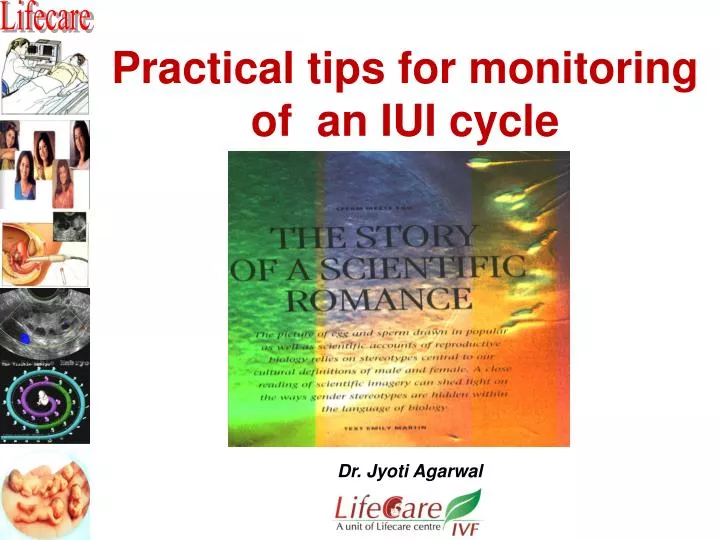
Practical tips for monitoring of an IUI cycle. Dr. Jyoti Agarwal. Introduction . Ovulation induction though sounds simple but there are many obstacles - Each patient behaves in a different fashion. - Variety of drugs and protocols are available.

E N D
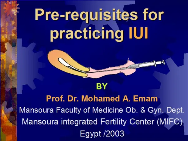
Practical tips for monitoring of an IUI cycle Dr. Jyoti Agarwal
Introduction • Ovulation induction though sounds simple but there are many obstacles - Each patient behaves in a different fashion. - Variety of drugs and protocols are available. • Every center has its own pattern of COH but the basic concept of monitoring remains the same.
Who should monitor? Why add to the burden ? Do it yourself
“Vision is the art of seeing invisible ”Jonathan swift • It is difficult to think of managing an infertile couple without resorting to this versatile and easy to use technology. • All the modalities of ultrasound ranging from basic black and white to the most complex , real time 3D and colour doppler have a role to play in managing these infertile patients .
Five Reasons To Monitor To evaluate if the dose being used is optimal To adjust the dose of the drug as some patients are hyper responsive and some are poor responders. To find the optimal time for inducing ovulation To time IUI To avoid excessive stimulation , to prevent OHSS and multiple pregnancy All patients to be monitored
Monitoring Should Be • Easy • Reliable • Patient friendly • Not expensive • Can be done by self
How to monitor ? • BY E 2 ALONE • BY ULTRASOUND ALONE • BY BOTH • BY COLOR POWER DOPPLER • BY OTHER HORMONES MINIMUM MONITORING
Monitoring Ultrasound states the morphological growth of the follicles Hormones indicates the functional activity of the follicles TVS is the accepted method by all ART centers.
Why TVS ? • Simple • Easy • Reproducible • Reliable • Cheap • Patient friendly
An transvaginal probe is an extension of clinician’s fingers ‘ marrying palpation with imaging ‘
Importance of D -2 scan • Antral follicle count • To rule out any cyst.( > 3 cm) • Endometrial shedding • Or any other pelvic pathology We expect normal sized ovaries with very small follicles (3—5 mm in diameter) Follicular size is measured by taking mean of 2 or 3 largest perpendicular diameters of each follicle .
Ultrasound follicular monitoring Serial USG follicular monitoring is started from day 7 or 8 of the cycle But in case of gonadotrophins we start scanning from 6th day of stimulation.
Assessing the follicular maturity • The follicles normally grow at a rate of 2- 3 mm / day in a stimulated cycle. • Definitive size of the follicle which confirms the maturity of oocytes is still controversial. • A follicle measuring 18—20 mm has been found to contain a mature oocyte.
Corelation with serum oestradiol levels • Plasma estradiol levels correlates closely with the stage of development of the dominant follicle • Serum estradiol levels >200 pg / ml on day 8 of stimulation indicates adequate dose of gonadotropins. Ultrasound monitoring has totally replaced estradiol monitoring in most centers.
Predicting the risk of OHSS If there are more than 4 follicles larger than 16 mm or more than 8 follicles larger than 12 mm It is best not to give hCG so as to prevent OHSS and high order multiple births. In case of doubt do serum estradiol levels Estradiol levels of > 1500 – 2000 pg/ml indicates risk of OHSS and is advisable to withhold hCG trigger.
Follicular doppler flow studies • A mature follicle shows vascularity in atleast ¾th of the follicular circumference and • PSV is 10 cm/sec. • At this time LH surge starts and • This is the right time to give hCG trigger
Perifollicular vascularisation Grade 1 : < 10% Grade 2 : 10-25% Grade 3 : 25-50% Grade 4 : > 50%
Predictors of poor ovarian response are : • Ovarian volume <3 cc • < 3 antral follicles • Ovarian RI > 0.6 • Ovarian PSV < 5 cm / sec • Stromal flow index < 11 • Suggest poor ovarian response & • Higher doses of gonadotropins will be required for stimulation.
ENDOMETRIAL EVALUATION Clear association between endometrial growth and the circulating estrogen & progesterone levels.
Endocrine implantation ET – 8 – 14 mm BEST ENDOMETRIUM ON THE DAY OF HCG TRIGGER ET > 16 mm or < 7mm Is not associated with good prognosis
Proliferative phase : 4- 7 mm • Periovulatory period : 6-10 mm • Secretory phase : 8-12 mm • Postmenopausal pd. : < 4 mm Thickest part of the endometrium should be measured
D-2 Can show • anechoic collection of blood. • thick echogenic endometrial echo . • a very thin endometrium 1-3 mm thick.
D3-7 • Increase in oestrogenic biosynthesis leads to stimulation and growth of endometrial glands and stroma. • Double line endometrium is seen which is usually < 6 mm.
D-7 onwards • Proliferative endometrium continues to grow in size and thickens and is seen as a triple layer or triple line. • Middle layer echogenic—Lumen
In Periovulatory Phase Triple line progressively becomes thicker, homogenous and hyperechoic
Endometrial evaluation Conception rates according to zones of vascularity • Zone 1 5.2 % • Zone 2 28 % • Zone 3 52 % • Zone 4 74%
Uterine Artery Doppler The chance for pregnancy is almost zero if the PI is more than 3.019 on the day of hCG administration Patients who get pregnant have a lower RI (0.53 vs 0.64)
Cervix and follicular monitoring On D – 13 scan Good cervical mucus • E2 > 100 pg • 2 follicles • ET 7-8 mm
Ovulation trigger The end point of any ovulation induction protocol is to indentify the best time for triggering ovulation. most crucial step In a gonadotrophinIn clomiphene Leading follicle isLeading follicle is 18 – 20 mm in diameter. 20 – 22 mm in size
hCG timing ALWAYS TIME HCG WITH FOLLICLE SIZE
Ovulation to be confirmed by • Disappearance of the follicle • Presence of free fluid in the cul-de-sac. • Presence of hyperechoic , smooth secretary endometrium.
Timing of insemination IUI is done 36 - 38 hrs. after hCG injection
Premature LH surge • Premature LH surge is known to occur in approx 15-25 % of patients once the leading follicle is 16 mm. • Urinary LH kits are available to detect LH surge. A blood level of >10 IU /L correlates with the LH surge
Premature LH surge • If an LH surge is detected , injection hCG is given immediately. • The hCG injection is required to supplement the LH secreted by the body as it is not adequate enoughto induce the final maturational changes in all the follicles . IUI is done 24 hrs after the LH surge
To conclude “ In the hands of experienced operators , ultrasound and ultrasound alone suffices for cycle monitoring .” • NEED OF EXTENSIVE HORMONAL • MONITORING IS NO LONGER NEEDED
All The Best to all of you to design your own Minimal Monitoring Protocol THANK YOU FOR HEARING ME OUT