Download
1 / 27
320 likes | 710 Views
Transfusion Reactions. NICHOLAS TSU, M.D. Objectives. Transfusion statistics and basics Types Diagnosis Treatment. Transfusion Statistics. Transfusions in 2004 14.2 million units of packed red blood cells (PRBC’s) 9.9 million units of platelets (84% apheresis units)

E N D
Transfusion Reactions NICHOLAS TSU, M.D.
Objectives • Transfusion statistics and basics • Types • Diagnosis • Treatment
Transfusion Statistics • Transfusions in 2004 • 14.2 million units of packed red blood cells (PRBC’s) • 9.9 million units of platelets (84% apheresis units) • 4.1 million units of fresh-frozen plasma (FFP) • Approximately 40% of all transfused units administered by anesthesia personnel
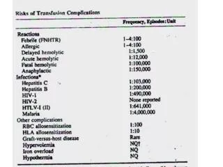
Transfusion Risks • Infectious • Viral • Bacterial • Noninfectious • Reaction to RBC Antigens • Acute Hemolytic Transfusion Reactions (AHTR) • Delayed Hemolytic Transfusion Reactions (DHTR) • Reactions to Donor Proteins • Minor Allergic Reactions • Anaphylactic Reactions • White Cell-Related Transfusion Reactions • Febrile Reactions • Transfusion-Related Acute Lung Injury (TRALI)
Bacterial Infections • Incidence of sepsis much greater in platelets • Platelets stored at room temperature • Decreased risk with apheresis platelets • Most common organisms • Staphyloccos aureus • Klebsiella pneumoniae • Serratia marcescens • Staphyloccos epidermdidis • Yersinia enterocolitica
Bacterial Sources • Donor skin flora • Donor bacteremia • Contamination from • Collection • Processing • Storage
Signs and Symptoms of Bacterial Infection • Fevers • Chills • Tacycardia • Dyspnea • Emesis • Shock • DIC • Acute renal failure
Diagnosis and Treatment • Stop transfusion • Obtain blood cultures • Treat with broad spectrum antibiotics • Notify the blood bank immediately • Prevent other units from same donor being transfused
Acute Hemolytic Transfusion Reactions • Most hazardous against foreign RBC’s • Hemolysis of donor RBC’s can lead to ARF & DIC • Mortality rate is 2% • Leading cause is clerical error
Acute Hemolytic Transfusion Reactions • Over 300 antigens on human RBC’s • Most common antibodies that fix complement • A, B, Kell, Kidd, Duffy • Rh antibodies do not fix complement but can cause serious hemolysis
AHTR Pathophysiology • Antibodies and complement in recipient plasma attack antigens on donor RBC’s causing hemolysis • Antigen-antibody complexes activate Hageman factor (factor XII) producing bradykinin leading to capillary permeability and hypotension • Complement system releases histamine and serotonin from mast cells resulting in bronchospasm • 30-50% of patients will develop DIC
AHTR Pathophysiology • Hemolysis releases hemoglobin (Hb) • Hb binds to haptoglobin and albumin initially • Will circulate unbound until excreted by kidneys • Renal damage causes • Hypotension 2/2 systemic hypotension and renal vasoconstriction • Free Hb form acid hematin damaging renal tubules • Antigen-antibody complexes may deposit in glomeruli
Signs and Symptoms • Fever • Chills • Nausea and vomiting • Diarrhea • Rigors • Hypotension and tachycardia (bradykinin) • Flushed and dyspneic (histamine) • Chest and back pain (cytokine release) • Headache • Feeling of impending doom • Hemoglobinuria eventually oliguria
Diagnosis • Stop transfusion • Recheck patient and unit labeling • Examine centrifuged plasma sample for pinkish discoloration representing free Hb • Hemolysis should be assumed to be hemolytic transfusion reaction until proven otherwise • Notify blood bank • Aseptically seal unit and return • Coombs test • Examines recipient RBC’s for presence of surface immunoglobulins and complement
Treatment • Maintain systemic blood pressure • Deliver volume • Pressors • Inotropes • Preserve Renal function and urine output • Administering fluids • Diuretics (mannitol or furosemide) • Sodium bicarb to alkalinize urine • Prevent DIC • No specific therapy • Prevent hypotension and support cardiac output • Decreases stasis
Delayed Hemolytic Transfusion Reactions • Compatible RBC’s are rapidly eliminated within days • Typically due to donor RBC antigen to which recipient has been previously exposed via transfusion or pregnancy • Over time antibody levels fall too low to be detected • With re-exposure anamnestic response results in antibodies and lysis of foreign RBC’s • Coated RBC’s are sequestered extravascularly (spleen and reticuloendothelial system) and lysed
Diagnosis and Treatment • Usually detected in the first or second week • Low-grade fever • Increased indirect bilirubin • Unexplained reduction in Hb • Decreased serum haptoglobin • Confirmed by positive Coomb’s test • Resolves as transfused cells are removed • Monitor Hb • Maintain hydration • Re-transfuse if necessary
Minor Allergic Reactions • Allergic reactions to proteins in donor plasma cause urticarial reactions in 0.5 to 4% of all transfusions • Most frequent in FFP or platelets • Itching, swelling, rash • Treat with diphenhydramine
Anaphylactic Reactions • Seen typically in pt’s with hereditary IgA deficiency • Previously sensitized during pregnancy or exposed to blood with foreign IgA • Dyspnea, bronchospasm, angioedema, hypotension • Discontinue transfusion • Administer epinephrine and methylprednisolone
Febrile Reactions • Pt’s who receive multiple transfusions of RBC’s will develop human leukocyte antigens (HLA) • On subsequent RBC transfusions antibodies attack donor leukocytes causing febrile reactions • Occur in up to 2% of platelet, FFP, and RBC transfusions • Increase in temperature of more than 1 degree C with 4 hours of transfusion • Defervesces within 48 hours • Occasional chills, dyspnea, anxiety, headache, myalgia • Treat with acetaminophen • Differentiate with direct Coomb’s test
Transfusion-Related Acute Lung Injury • TRALI is a noncardiogenic form of pulmonary edema occurring after blood product administration • Associated with all plasma-containing components • Estimated at 1:1271 to 1:5000 transfusions • Mortality of at least 5%
Transfusion-Related Acute Lung Injury • Occurs when mediators present in the plasma of donor blood activates leukocytes in the host • Activated leukocytes are sequestered by the lungs • Leukocyte mediators are released and cause increased capillary permeability and endothelial damage • “two hit theory” • Trauma, surgery, sepsis may first “prime” native granulocytes causing surface adhesion sites resulting lung sequestration • Biologically active mediators that are breakdown products from cellular elements in blood products activate sequestered leukocytes
Signs and Symptoms • Within 6 hours of transfusion • Dspnea • Chills • Fever • Noncardiogenic pulmonary edema/bilateral pulmonary infiltrates • Hypotension/hypertension may occur
Diagnostic Criteria • Acute onset of hypoxemia (within 6 hours of conclusion of transfusion) • Bilateral CXR infiltrates consistent with ALI • Absence of evidence of left atrial hypertension • Absence of temporally related causes of ALI
Treatment • Largely supportive • Transfusion should be stopped if recognized in time • Supplemental oxygen and ventilation support provided if necessary • Use low tidal volume settings like in ARDS • No diuretics • Glucocorticoids have been administered but no evidence supporting their administration