Download
1 / 30
300 likes | 418 Views
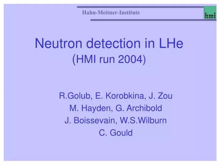
Neutron detection in LHe ( HMI run 2004). R.Golub, E. Korobkina, J. Zou M. Hayden, G. Archibold J. Boissevain, W.S.Wilburn C. Gould. Layout of the neutron beam. Li shield. Li Beam stop when needed. Lead shield. Beam stop. Beam monitor. Lead shield. Bi filter.

E N D
Neutron detection in LHe(HMI run 2004) R.Golub, E. Korobkina, J. Zou M. Hayden, G. Archibold J. Boissevain, W.S.Wilburn C. Gould
Layout of the neutron beam Li shield Li Beam stop when needed Lead shield Beam stop Beam monitor Lead shield Bi filter Cross section of the full beam was 1 cm2,intensity ≈ 106 n/cm2/s before Bi The distance from the polarizer to the light window was ≈ 120 cm Guide Polarizer Collimator, Li Chopper and neutron guide of SANS Light guide 33 cm
Layout of the cell and light collecting system Light window of the cell OD of the cell =5 cm, ID = 4.4 cm The distance from the neutron beam to the light window, L, was L≈2 OD 77K 4K 300K Acrylic cell 22 cm PMT C2 Light guide 33 cm Neutron beam Al wall, 2 mm thick Reflector - Al foil Cell Cross section
View looking `upstream’ (toward the reactor) Cryostat Beam Stop
t PMT oscilloscope Collimation cell neutrons light guides PMT delay line
Cell, as viewed through PMT window free liquid surface
What was studied We have studied at different temperatures • Empty cell and Neutron guide hall background 2. Cell filled with Natural He - 90 mK, 300 mK, 700 mK, 1400 mK Full beam opened and closed with Li. 3. Cell with additional He-3 (about 100 ppm) • 90 mK, 300 mK, 500 mK, 650 mK, 1500 mK, Full beam opened and closed with Li. • Small beam opened, 90 mK, 120 mK, 300 mK, 500 mK, 1500 mK, 1900 mK, 90 mK with and without coincidence, 10 mV LDL and 15 mV LDL • Ultra Small beam opened, 90 mK, 1500 mK, 1900 mK Data were taken in two modes - Count Rate, where 10 ms intervals were recorded, time scale = 10 or 1 ns/point - Sequence, where only coincided events and after pulses during 20 mks were recorded, time scale = 1 ns/point We have used LeCroy oscilloscope with a time scale down to 125 ps per point
Count Rate data evaluation Our data allowed us to compare Count Rate data evaluation with and without coincidence. Without coincidence is a real count rate of the detectors that is a sum of the background from acrylic light guides and the cell’s signals. Coincidence mode is a great tool to get read of the background pulses from the light guides.
First big run; cell is empty, PMT HV=2kV, time scale 1 ns/point to see small pulses. A single photon (SP) peak and gamma are well separated. Gamma peak = 3 SP. Empty cell, no coincidence
Beam On, Li in the beam PMT pulse area spectra at different temperatures when the beam is stopped by Li rubber . • Left - PMT C2, 500 mK, 300 mK with He-3 and 98 mK, Natural Helium • Right - PMT C4, 500 mK, 1500 mK with He-3 and 98 mK, Natural Helium
Full beam and Small beam • Full beam shows too high intensity of single pulses. • We made the beam smaller • Count Rate over the neutron peak range *************************************************** • C4 Small 4906 • C4 Li 1733 • C2 Small 4632 • C2 Li 1472 Neutrons 3000 sec-1 *************************************************** • C4 Full 19067 • C4 Nat He 4710.8 • C2 Full 18605 • C2 Nat He 4134.41 Hz Neutrons 14 000 sec-1 ***************************************************
Small and Ultra Small beams • The main set of data was taken for the Small Beam. • In addition, we took several runs with a beam even smaller to get more close to EDM conditions • Ultra Small Beam • Count Rate over the neutron peak range *********************************************** • C4 USB 2927 • C4 Li 1733 • C2 USB 2714 • C2 Li 1472 Neutrons 1000 sec-1 *********************************************** Ratio Gamma’s/Neutrons 3/2
Full Beam, temperatures PMT spectra at different temperatures. 90 mk 350 mK 630 mK 1500 mK 90 mK were measured at two slightly different vertical scales. Setup 1 was used for all Natural He runs Setup 4 - for all He-3 runs
Count Rate in coincidence, Nat.He Here count rates for channels C2 and C4 were evaluated in coincidence. Note that here the Low Discrimination Level (LDL) is below a Single Photon amplitude. Practically, here only presence of any pulse is a trigger. From 0.2 nVsec up to 1.5 nVsec count rate is: • C2 ,C4 Nat He, Full beam ~ 2200 Hz • C2,C4 Nat He Full beam Li ~ 550 Hz
Beam with Li and Full beam in Natural He • Count Rate (s-1) integrated over the neutron peak range [0.2,1.5] nVs • C2 Li 1471 • C2 Nat He 4134.41 • C4 Li 1733 • C4 Nat He 4710.8 • High energy tail is result of the beam entering the cell
PMT HV=1.9kV, time scale 10 ns/point (for count rate mode) to see larger neutron’s pulses. A single photon (SP) peak and gamma are not separated. Gamma spectra show no temperature dependence. Count rate is very stable. He-3 in the cell, Full Beam with Li, no coincidence
Neutron Peak, C2 and C4 • Neutron peak estimated from the Full beam data
Count Rate in coincidence, He-3, Ultra Small beam & Full Beam, Nat.He
Neutron Peak in coincidence, He-3, Ultra Small beam - Full Beam with Li, Nat.HeCount rate is close to expected one for EDM
C2+C4, LDL=15 mV - the same conditions as in the sequence mode
Sequence data evaluation Sequence data were recorded with LDL = 15 mV and coincidence time = 20 ns, during 20 mks after a trigger. The trigger position was 1.5 mks. For 2D plot (presented here) we have integrated area of the main pulse (over 100 ns) and counted after pulses during 6 mks after the main pulse. The first 1 mks and last 10 mks were used to estimate the afterpulse background. We used sum of pulse areas and numbers of afterpulses of both PMT : C2 and C4
Single Photon Peak 2D temperature dependence 1900 mK 300 mK
Single Photon Peak 2D Small beam & Full Beam with Li
Rough Comparison with the NIST cell HMI beam position Our position is at about 50% of the light collection efficiency. The far end of EDM cell of proposal is at about 30% that implies a neutron peak position at 0.6 nVs 0r 12 single photons (at HMI we have the neutron peak at 20 SP ) Position of a far end of the cell from the EDM proposal Light Guide
EDM cell • Probably, light collection can be made from both sides. • One side with higher efficiency than another • If no light collection from the opposite side, at least, the light guide must be split into 2 parts
Summary • Our data allowed us to compare Count Rate data evaluation with and without coincidence. • We see that coincidence mode is a great tool to get read of the background pulses from the light guides. • In coincidence we can see clear 400 Hz peak of neutrons with the beam 104 n/sec in the cell in 1D plot. • 2D plot with afterpulses shows even better separation while not as good as at higher temperatures. • The single PMT amplitude of the neutron beam is 20 single photons even under our not very optimized conditions(cell mis alignment! ). • Our data can be used to simulate quite precisely EDM data. This is only start of our data evaluation!