Download
1 / 68
680 likes | 709 Views
Learn about various types of microscopy, including light and electron microscopes, staining techniques, and principles such as contrast and resolution. Understand why scientists use metric units instead of English units.

E N D
Units of Measurement • Tell Me Why • Why do scientists use metric rather than English units?
Microscopy • Microscopy: the use of light or electrons to magnify objects • Science of microscopy begun by Antoni van Leeuwenhoek • Various types of light and electron microscopes
Microscopy • General Principles of Microscopy • Wavelength of radiation • Magnification • Resolution • Contrast
Figure 4.2 Light refraction and image magnification by a convex glass lens. Light Air Glass Focal point Inverted, reversed, and enlarged image Specimen Convex lens
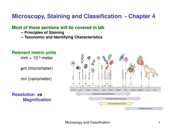
Figure 4.3 The limits of resolution (and some representative objects within those ranges) of the human eye and of various types of microscopes.
Microscopy • General Principles of Microscopy • Contrast • Differences in intensity between two objects or between an object and its background • Important in determining resolution • Staining increases contrast • Use of light that is in phase increases contrast
Microscopy • Light Microscopy • Bright-field microscopes • Simple • Contain a single magnifying lens • Similar to magnifying glass • Leeuwenhoek used simple microscope to observe microorganisms
Microscopy • Light Microscopy • Bright-field microscopes • Compound • Series of lenses for magnification • Light passes through specimen into objective lens • Oil immersion lens increases resolution • Have one or two ocular lenses • Total magnification = magnification of objective lens x magnification of ocular lens • Most have condenser lens (direct light through specimen)
Figure 4.4 A bright-field, compound light microscope. Line of vision Ocular lens Remagnifies the image formed by the objective lens Ocular lens Body Transmits the image from the objective lens to the ocular lens using prisms Path of light Prism Arm Objective lenses Primary lenses that magnify the specimen Body Objective lenses Stage Holds the microscope slide in position Specimen Condenser Focuses light through specimen Condenser lenses Diaphragm Controls the amount of light entering the condenser Illuminator Illuminator Light source Coarse focusing knob Moves the stage up and down to focus the image Fine focusing knob Base
Figure 4.5 The effect of immersion oil on resolution. Microscope objective Microscope objective Lenses More light enters lens Refracted light rays lost to lens Immersion oil Glass cover slip Glass cover slip Slide Slide Specimen Light source Light source Without immersion oil With immersion oil
Microscopy • Light Microscopy • Dark-field microscopes • Best for observing pale objects • Only light rays scattered by specimen enter objective lens • Specimen appears light against dark background • Increases contrast and enables observation of more details
Figure 4.6 The light path in a dark-field microscope. Objective Light refracted by specimen Light unrefracted by specimen Specimen Condenser Dark-field stop Dark-field stop
Microscopy • Light Microscopy • Phase microscopes • Used to examine living organisms or specimens that would be damaged/altered by attaching them to slides or staining • Light rays in phase produce brighter image, whereas light rays out of phase produce darker image • Contrast is created because light waves are out of phase • Two types • Phase-contrast microscope • Differential interference contrast microscope
Figure 4.7 Principles of phase microscopy. Rays in phase Rays out of phase Phase plate Bacterium Deviated ray is now 1/2 wavelength out of phase. Ray deviated by specimen is 1/4 wavelength out of phase.
Figure 4.8 Four kinds of light microscopy. Nucleus Bacterium Dark field Bright field Phase contrast Nomarski
Microscopy • Light Microscopy • Fluorescent microscopes • Direct UV light source at specimen • Specimen radiates energy back as a longer, visible wavelength • UV light increases resolution and contrast • Some cells are naturally fluorescent; others must be stained • Used in immunofluorescence to identify pathogens and to make visible a variety of proteins
Figure 4.10 Immunofluorescence. Fluorescent dye Antibodies Antibodies carrying dye Bacterium Cell-surface antigens Bacterial cell with bound antibodies carrying dye
Microscopy • Light Microscopy • Confocal microscopes • Use UV lasers to illuminate fluorescent chemicals in a single plane • Resolution increased because emitted light passes through pinhole aperture • Each image is "optical slice" through specimen • Computer constructs 3-D image from digitized images
Microscopy • Electron Microscopy • Light microscopes cannot resolve structures closer than200 nm • Electron microscopes have greater resolving power and magnification • Magnifies objects 10,000x to 100,000x • Detailed views of bacteria, viruses, internal cellular structures, molecules, and large atoms • Two types • Transmission electron microscopes • Scanning electron microscopes
Figure 4.11 A transmission electron microscope (TEM). Light microscope (upside down) Column of transmission electron microscope Lamp Electron gun Condenser lens Condenser lens (magnet) Specimen Specimen Objective lens Objective lens (magnet) Projector lens (magnet) Eyepiece Final image on fluorescent screen Final image seen by eye
Figure 4.12 Scanning electron microscope (SEM). Electron gun Magnetic lenses Beam deflector coil Scanning circuit Primary electrons Secondary electrons Photo- multiplier Specimen Monitor Detector Specimen holder Vacuum system
Microscopy • Probe Microscopy • Magnifies more than 100 million times • Two types • Scanning tunneling microscopes • Atomic force microscopes
Microscopy • Probe Microscopy • Scanning tunneling microscopes • Passes metallic probe above specimen surface • Measures the electron flow (tunneling current) to and from the probe and the specimen's surface • Atomic force microscopes • Passes probe lightly on the specimen surface • Deflection of laser beam translated into atomic topography
Figure 4.14 Probe microscopy. Enzyme DNA
Microscopy • Tell Me Why • Why is magnification high but color absent in an unretouched electron micrograph?
Staining • Most microorganisms are difficult to view by bright-field microscopy • Coloring specimen with stain increases contrast and resolution • Specimens must be prepared for staining
Staining • Principles of Staining • Dyes used as stains are usually salts • Chromophore is the colored portion of the dye • Acidic dyes stain alkaline structures • Basic dyes stain acidic structures • More common because most cells are negatively charged
Staining • Simple stains—composed of single dye • Differential stains—use more than one dye • Gram stain • Acid-fast stain • Endospore stain • Histological stains • Special stains—reveal specific structures • Negative (capsule) stain • Flagellar stain
Figure 4.19 Schaeffer-Fulton endospore stain of Bacillus anthracis.
Staining • Differential Stains • Histological stains • Two common stains used for histological specimens • Gomori methenamine silver (GMS) stain • Hematoxylin and eosin (HE) stain
Figure 4.20 Negative (capsule) stain of Klebsiella pneumoniae. Bacterium Capsule Background stain
Staining • Staining for Electron Microscopy • Chemicals containing heavy metals are used for transmission electron microscopy • Stains may bind molecules in specimens or the background
Microscopy • Tell Me Why • Why is a Gram-negative bacterium colorless but a Gram-positive bacterium purple after it is rinsed with decolorizer?