Download
1 / 26
260 likes | 273 Views
11 The Muscular System. An Introduction to the Muscular System. Learning Outcomes 11-1 Describe the arrangement of fascicles in the various types of muscles, and explain the resulting functional differences.

E N D
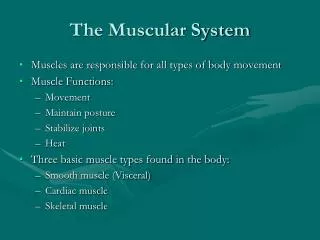
11 The Muscular System
An Introduction to the Muscular System Learning Outcomes 11-1 Describe the arrangement of fascicles in the various types of muscles, and explain the resulting functional differences. 11-2Describe the classes of levers, and explain how they make muscles more efficient. 11-3 Predict the actions of a muscle on the basis of its origin and insertion, and explain how muscles interact to produce or oppose movements. 11-4 Explain how the name of a muscle can help identify its location, appearance, or function. 11-5 Identify the principal axial muscles of the body, plus their origins, insertions, actions, and innervation. 11-6 Identify the principal appendicular muscles of the body, 11-7 Identify age-related changes of the muscular system. 11-8 Explain the functional relationship between the muscular system and other body systems, and explain the role of exercise in producing various responses in other body systems.
An Introduction to the Muscular System The Muscular System (only the skeletal muscles) Muscle Organization and Function Muscle organization affects power, range, and speed of muscle movement Fascicles (muscle cells (fibers) are organized in bundles (fascicles) Classification of Skeletal Muscles By the way fascicles are organized By relationships of fascicles to tendons Organization of Skeletal Muscle Fibers Four patterns of fascicle organization Parallel Pennate Convergent Circular
11-1 Fascicle Arrangement Parallel Muscles Fibers parallel to the long axis of muscle For example,biceps brachii Depends on total number of myofibrils Directly relates to cross section of muscle 1 in.2 (6.45 cm2) of cross section develops 50 lb (23 kg) of tension
11-1 Fascicle Arrangement Convergent Muscles A broad area converges on attachment site (tendon, aponeurosis, or raphe) Muscle fibers pull in different directions, depending on stimulation For example,pectoralis muscles
11-1 Fascicle Arrangement Pennate Muscles Form an angle with the tendon Develop more tension than parallel muscles Unipennate Fibers on one side of tendon For example, extensor digitorum Bipennate Fibers on both sides of tendon For example, rectus femoris Multipennate Tendon branches within muscle For example, deltoid
11-1 Fascicle Arrangement Circular Muscles Also called sphincters Open and close to guard entrances of body For example,orbicularis oris muscle of the mouth
11-2 Levers Skeletal Motion Skeletal muscles attach to skeleton, produce motion Type of muscle attachment affects power, range, and speed of muscle movement Levers (a rigid piece turning about an axis and used for transmitting and changing force and motion). Mechanically, each bone is a lever (a rigid, moving structure) And each joint a fulcrum (a fixed point) Muscles provide applied force (AF) Required to overcome load (L)
11-2 Levers Function of a Lever To change: Direction of an AF Distance and speed of movement produced by an AF Effective strength of an AF The Three Classes of Levers Depend on the relationship between applied force, fulcrum, and resistance • First-class lever • Second-class lever • Third-class lever
1 Which type of lever prodouces the most force?
11-2 Levers First-Class Lever Seesaw or teeter-totter is an example Center fulcrum between applied force and load Force and load are balanced
11-2 Levers Second-Class Lever Wheelbarrow is an example Center resistance between applied force and fulcrum A small force moves a large weight
11-2 Levers Third-Class Lever Most common levers in the body Center applied force between load and fulcrum Greater force moves smaller load Maximizes speed and distance traveled
11-3 Muscle Attachments to Other Tissues Origins and Insertions Muscles have one fixed point of attachment (origin) And one moving point of attachment (insertion) Most muscles originate or insert on the skeleton Origin is usually proximal to insertion Actions Movements produced by muscle contraction Body movements For example, flexion, extension, adduction, etc. Described in terms of bone, joint, or region
11-3 Muscle Attachments to Other Tissues Muscle Interactions Muscles work in groups to maximize efficiency Smaller muscles reach maximum tension first, followed by larger, primary muscles Muscle Terminology Based on Function Agonist (or prime mover)—Produces a particular movement Antagonist—Opposes movement of a particular agonist Synergist—Smaller muscle that assists a larger agonist Helps start motion or stabilize origin of agonist (fixator)
11-3 Muscle Attachments to Other Tissues Muscle Opposition Agonists and antagonists work in pairs When one contracts, the other stretches Such as flexors–extensors, abductors–adductors, etc. Names of Skeletal Muscles Correct names of muscles include the term muscle Exceptions: Platysma (covers anterior part of neck) Diaphragm (divides thoracic and abdominopelvic cavities)
Give an example of a muscle names for each of the following characteristics: direction of fibers, shape, action, size, origin and insertion, location, and number of tendons of origin.
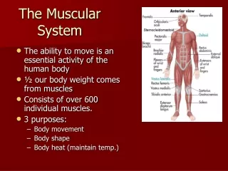
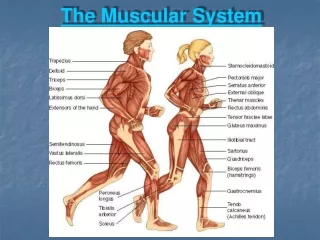
11-4 Naming Skeletal Muscles Divisions of the Muscular System Axial muscles Position head and spinal column Move rib cage 60% of skeletal muscles Appendicular muscles Support pectoral and pelvic girdles Support limbs 40% of skeletal muscles
TheAxial Muscles • Divisions based on location and function • Muscles of the head and neck • Muscles of the vertebral column • Oblique and rectus muscles • Muscles of the pelvic floor The Appendicular Muscles Position and stabilize pectoral and pelvic girdles Move upper and lower limbs Two divisions of appendicular muscles Muscles of the shoulders and upper limbs Muscles of the pelvis and lower limbs
11-7 Effects of Aging on the Muscular System Effects of Aging Skeletal muscle fibers become smaller in diameter Skeletal muscles become less elastic Develop increasing amounts of fibrous tissue (fibrosis) Decreased tolerance for exercise Decreased ability to recover from muscular injuries
11-8 Muscular System Integration Cardiovascular System Delivers oxygen and fuel Removes carbon dioxide and wastes Respiratory System Responds to oxygen demand of muscles Integumentary System Disperses heat from muscle activity Nervous and Endocrine Systems Direct responses of all systems
BIOL 2401 1 An electrical signal (nerve impulse) moves along the nerve toward the nerve ending. Stored within the nerve ending are vesicles filled with ACh. 2 The nerve impulse causes the vesicles to release the ACh into the space between the nerve ending and the muscle membrane. 3 ACh diffuses across the space and binds to the receptor sites on the muscle membrane. 4 The ACh stimulates the receptors and causes an electrical signal to develop along the muscle membrane. The ACh then unbinds the receptor site and is immediately destroyed by AChE. The free-binding sites are then ready for additional Ach when the nerve is stimulated again. Abnormal Functioning at the NMJ
BIOL 2401 Match the examples of abnormalfunctioning at the NMJ. Picture 1 Myasthenia gravis Picture 2 Neuromuscular blockade by curare Picture 3 Infection by Clostricium botulinum Picture 4 Infection by Clostridium tetani