Download
1 / 31
330 likes | 884 Views
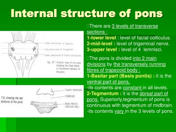
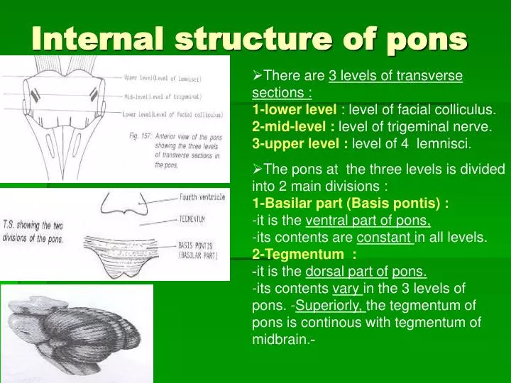
Internal structure of pons. There are 3 levels of transverse sections : 1-lower level : level of facial colliculus. 2-mid-level : level of trigeminal nerve. 3-upper level : level of 4 lemnisci.

E N D
Internal structure of pons • There are 3 levels of transversesections :1-lower level : level of facial colliculus. 2-mid-level : level of trigeminal nerve. 3-upper level : level of 4 lemnisci. • The pons at the three levels is divided into 2 main divisions : 1-Basilar part (Basis pontis) : -it is the ventral part of pons, -its contents are constant in all levels. 2-Tegmentum : -it is the dorsal part ofpons. -its contents vary in the 3 levels of pons.-Superiorly, the tegmentum of pons is continous with tegmentum of midbrain.-
Ventral (basilar) part of pons in all levels: • It is marked by numerous transverse pontocerebellar fibres which arise from pontine nuclei buried in its substance. • These transverse Pontocerebellar fibres cross the midline to pass to contralateral cerebellar hemisphere forming middle cerebellarpeduncle (brachium pontis), where trigeminal N. (V) pierce it. . Mid-pons at level of trigminal nerve. Caudal pons at level of facial colliculus
Ventral (Basilar)part of pons in all levels: • Corticospinal fibres & corticobulbar (pyramidal tract) appear as small,separate bundles running longitudinally between fascicles of transverse pontine fibres. T.S of caudal pons at level of facial colliculus T.S of rostral pons at level of 4 lemnisci T.S of mid-pons at level of trigeminal nerve
Dorsal( tegmental) part of caudal pons at level of facial colliculus : 1-Trapezoid body consists of acoustic fibresresponsible for hearing, arising from cochlear nuclei (dorsal & ventral cochlear nuclei lying dorsal & ventral to inferiorcrebellar peduncle in the most lower part of pons). -These acoustic fibres crossing opposite side of pons forming decussation oftrapezoid body, then the axons ascend into rostral pons & midbrain as lateral lemniscus and terminate in inferior colliculus. -it lies in the anterior part of tegmentum. T.S of caudal pons T.S of caudal pons T.S of rostral pons
Dorsal( tegmental) part of caudal pons at level of facial colliculus : 2-Medial lemniscus:-(the most medial lemniscus) is marking the boundarybetween ventral & tegmental portions of pons. -It is a band of ascending fibres carrying proprioceptive & fine touch sensation from opposite side of body, via Gracile & Cuneate tracts,ending in P.L.V.N. of thalamus. T.S of caudal pons T.S of rostral pons
Dorsal (tegmental)part of caudal pons at level of facial colliculus : 3-Medial longitudinal fasciculus or bundle : -it is an association tract present close to midline , found throughout the brain stem,and descend into spinal cord. -It links vestibular nuclei with motor ocular nuclei, supplying extraocular muscles (oculomotor,trochlear & abducent nuclei) to serve corrdination of head and eye movements.
Dorsal (tegmental)part of caudal pons at level of facial colliculus : 4-Abducent nerve nucleus :site :in posterior aspect of caudal pons near floor of 4th ventricle. It is encircled by fibres of facial N., forming elevation in the the floor of 4th ventricle called facial colliculus. -Its efferent fibres :pass downwards traversing medial lemniscus & pyramidal T.bundles to emerge anteriorly at junctionbetween pons & medulla, supplying lateral rectus muscle 5-Facial motor nucleus :site : in posterior part of caudal pons.Its efferent fibres encircle abducent nucleus,then pass anterolaterally to emerge at the junction between pons &medulla,(supplying ms. of facial expression) T.S through caudal part of pons at the level of facial colliculus.
Dorsal (tegmental) part of caudal pons at level of facial colliculus : 6-Vestibular nuclei : they are 4 nuclei lie subjacent to vestibular area of floor of 4th ventricle. -they receive afferent fibres from the vestibular nerve. -they send efferent fibres as :vestibulo-ocular fibres through medial longitudinal fasciculus. 7-Spinal tract & nucleus of trigeminal nerve: lie on the anteromedial aspect of inferior cerebellar peduncle. -carrying pain &temperature sensationsfrom the face. -The axons of the cells of the nucleus cross to opposite side and ascend in trigeminallemniscus to end in thalamus. 8-Inferior cerebellar peduncle. T.S through caudal part of pons at the level of facial colliculus.
Dorsal (tegmental)part of mid-pons at level of trigeminal nerve : 1-Trigeminal motor nucleus : medial in position. its axons form the motor root of trigeminal N. which passes along mandibular N. (supplying muscles of mustication). 2-Trigeminal sensory nucleus : lateral in position. It receives afferent touch & pressure sensation from face. -It sends efferent fibres which cross to join trigeminal tract or lemniscus. 3-Superior cerebellar peduncle : lies posterolateral to motor nucleus of trigeminal nerve. 4-Medial longitudinal fasciculus, spinal lemniscus , medial lemniscus & trapezoid body. T.S of mid-pons a level of trigeminal N. .
Dorsal (tegmental) part of Rostral Pons at level of 4 lemnisci : 1-Superior cerebellar peduncle :lies in the rostral part of pons, forming lateral walls of 4th ventricle. -They are connected together by superior medullary velum which forms roof of 4th ventricle. T.S of rostral pons T.S of mid-pons at level of trigeminal nerve.
Dorsal (tegmental) part of Rostral Pons at level of 4 lemnisci : • Types of fibres in the S.C.P :(A) Afferent fibres :1-ventral spino-cerebellar tract : it carries proprioceptive impulsesfrom the limbs to cerebellum. 2-tecto-cerebellar tract : it carries auditory & visual impulses fromtectum of midbrain to cerebellum. (B)Efferent fibres :1-Dendato-rubral tract : it is concerned with coordination ofmovement. it ends in red nucleus (Extrapyramidal nucleus) in midbrain.2-Dentato-thalamic tract : from dentate nucleus of cerebellum to end in ventral lateral nucleus ofthalamus. T.S of rostral pons
Dorsal (tegmental) part of Rostral Pons at level of 4 lemnisci : 2-Lateral lemniscus : themost lateral lemniscus. it is a band of ascending fibres carryinghearing sensation from both ears ( mainly from opposite side), via acoustic Fs. Of cochlear nuclei, Ending inauditory area in temporal lobe. 3-Spinal lemniscus :justmedial to lateral lemniscus. it is a band of ascending Fs. Carryingpain, tempreture & crude touch from opposite side of body via spinothalamic tractEnding inP.L.V.N.of thalamus.
Dorsal (tegmental) part of Rostral Pons at level of 4 lemnisci : 4-Trigeminal lemniscus: just medial to spinal lemniscus.it is a band of ascending Fs.carrying pain, temp., touch & proprioception from opposite side of face & scalp, via sensory Fs. Of trigeminal N. ending inP.M.V.N.of thalamus. 5-Medial lemniscus : -(the most medial lemniscus) is marking the boundarybetween ventral & tegmental portions of pons. - it is a band of ascending fibres carrying proprioceptive & fine touch sensation from opposite side of body, via Gracile & Cuneate tracts,ending in P.L.V.N. of thalamus.
Examples for Questions of Pons : 1. All of these tracts are found in the pons EXCEPT: a.Corticospinal tract. b.Corticobulbar tract. c.Medial lemniscus. d.Medial longitudinal fasiculus. e.Gracile tract.
Pons : 2. Which nucleus is not lying in the tegmentum of pons ? a. Facial motor nucleus. b. Abducent motor nucleus. c. Inferior olivary nucleus. d. Cochlear nucleus. e. Vestibular nucleus. 3. The acoustic fibres ascend as : a. Medial lemniscus. b. Spinal lemniscus. c. Lateral lemniscus. d. Trigeminal lemniscus. e. Medial longitudinal tract.
Internal structure of Midbrain • The midbrain is divided into dorsal & ventral portions at the level of cerebral aqueduct.(A)Tectum :the smaller dorsal part behind aqueduct. It is composed of 4 rounded swellings (colliculi) : 1-2 superior colliculi :lower centers of vision.2-2 inferior colliculi : lower centers of hearing.(B)2 Cerebral peduncles :the larger ventral part in front of aqueduct. It consists of 3 parts :1-Crus cerebri (Basis pedunculi) :the most anterior part which consists entirely of pyramidal &cortico-pontine fibres.2-Substantia nigra : a thick lamina of grey matter formed of deeply pigmented nerve cells lying behind crus cerebri. It is anExtrapyramidal motor centre.3-Tegmentum :the post. part of cerebral peduncle. It contains ascending tract, decussation, nuclei, & reticular formation.
Caudal midbrain at level of Inferior colliculus 1-The inferior colliculus is a centreof hearing reflex which receives ascending auditory pathway ,which run in lateral lemniscus. -Its Efferent Fs. end in medial geniculate nucleus of thalamus, which projects to auditory cortex of temporal lobe. 2-The cerebral aqueduct runs ventral to colliculi, and surrounding by area of grey matter, the peri-aqueductal (or central grey ). 3-Trochlear nucleus : lies ventral to peri-aqueductal grey, its efferent Fs. cross to opposite side to emerge from back of midbrain, then turn forwards to reach base of brain to supply extraocular ms.(sup.oblique). 4-Mesencephalic nucleus of Trigeminal :-lies lateral to aqueduct of midbrain, at level of inferior&superior colliculi.Sensory nucleus receives proprioceptive sensation from ms.of mastication. s.
Auditory pathway & inferior colliculus of midbrain (reflex center of hearing) :
Caudal midbrain at level of Inferior colliculus 5-Medial longitudinal fasciculus : is a well defined bundle of association fibres lies on each side of median plane in midbrain tegmentum. -It extends throughout the brain stem, and descends into spinal cord. -It lies close to oculomotor, trochlear & abducent nuclei. -it receives fibres from vestibular nuclei.-it sends efferents to ocular motor nuclei-Its function:coordination of eye,and head & neckmovements.
Caudal midbrain at level of Inferior colliculus 6-Decussation of superior cerebellar peduncles(brachium conjunctivum) :the fibres of each peduncle cross to opposite side, forming decussation in the central part of tegmentum. 7-Medial lemniscus :it is a band of ascending Fs. carrying proprioceptive sensation from opposite side of body. -It is the upward continuation of gracile & cuneate tracts of opposite side. -It lies in tegmentum, posterior to substantia nigra. .
Caudal midbrain at level of Inferior colliculus 8-Substantia Nigra : It is a large extrapyramidal motor nucleus, lies at midbrain tegmentum.-It contains subdivision part, the pars compacta, which consists of pigmented, melanin-containing neurones that synthesize dopamine astheir transmitter. -Itproject to caudate nucleus+putamen of basal ganglia in the forebrain. --It has extra-pyramidal motor function,concerned with movements. -lesionof pars compacta leads to parkinson’s disease.It is due toabsence of dopamine into basal ganglia, this is manifested bya mask face, resting tremors, rigidity of muscles (more in flexors giving flexor attitude) anda shuffling gait. T.S.of caudal midbrain at level of inferior colliculus.
Caudal midbrain at level of Inferior colliculus 9-Crus Cerebri :lies ventral tosubstantia nigra. It consists entirely of descending cortical efferent Fs.-50% of crus consists of pyramidal tract consists of cortico-bulbar Fibres (end in motor cranial nerve nuclei of brain stem) +cortico-spinal fibres down to medullary pyramid and spinal cord. -on either side ofcorticobulbar & corticospinal fibres, crus cerebri contains cortico-pontine,temporo-pontine + fronto-pontine fibres-These Fs. arise from cerebral cortex and ends in pontine nuclei of ventral pons to pass via M.C.P into cerebellum,(cortico-ponto-cerebellar pathway ) to involve in coordination of movement. T.S.of caudal midbrain at level of inferior colliculus.
Rostral midbrain at the level of Superior Colliculus: • Superior colliculus : -lies in upper part of tectum of midbrain. -it is a centre of visual reflex. -Its main afferent Fs. are : Cortico-tectal Fs. arise from -visual cortex of occipital lobe. -frontal eye field of frontal lobe.-function : control movements of eyes + accomodation reflex. • Pretectal nucleus : It lies above the superior colliculus. -It receives the visual Fs. running in optic tract just rostral to superior colliculus. -it has connections with parasymp. nucleus of oculomotor N. (Edinger-Westphal nucleus) to control smooth ms. of eye (sphincter pupillae) and to mediate pupillary light reflex. .
Rostral midbrain at the level of Superior Colliculus: • Peri-aqueductal (central) grey. • Oculomotor nucleus : lies ventral to peri-aqueductal grey at level of superiorcolliculus of midbrain. -efferent Fs. emerge from the medialsurface of crus cerebri. as oculomotor nerve to supply extraocular ms. of eye (except S.O + L.R.). • Red nucleus : -it is a large mass of grey matter lies in tegmentum of rostral midbrain. -it has spinal extrapyramidal motor function. -It receives afferents from motor cortex & cerebellum (cortico-rubral & dentato-rubral F.). -it sends efferents to spinal cord as rubro-spinal tract.
Reticular formation • It is a gray matter extending throughout the length of brain stem, made up of , deeply placed nerve cells & fibres. • It has important functions for vital centers as respiratory & cardiovascular centres. • It has descending fibres,reticulospinal tractsthat influence muscle tone &posture. • It has ascending fibres,the reticularactivatingsystem (RAS),plays a role in consciousness and awake / sleep cycle.
Reticular formation • Raphe nuclei : are a group of midline nuclei that extend throughout the length of brain stem. -they are serotonergic nuclei (their transmitter is serotonine). -Their ascending fibres to forebrain are involved in neural mechanisms regulating sleep. -Descending fibres to the spinal cord are involved in modulation of nociceptivemechanisms. • Locus coeruleus : is a group of pigmented neurones that lies in brain stem tegmentum of caudalmidbrain & rostral pons. -it is noradrenergic cell group. -it has ascending fibres to cerebellum, thalamus,hypothalamus, limbic system and cerebral cortex. -its descending fibres project to brain stem & spinal cord. -involved in neural mechanisms regulating sleep. Raphe nuclei Locus coeruleus
Brain stem lesions : • A unilateral brain stem lesion : caused by stroke,tumour or multiple sclerosis causes : 1-epsilateral cranial nerve dysfunction + contralateral spastic hemiparesis. 2-hyperreflexia & an extensor plantar response (upper motor neurone lesion). 3-contalateral hemisensory loss. 4-ipsilateral incoordination. 5-it can affect eye movements through demyelination of medial longitudinalfasciculus, producing internuclear ophthalmoplegia which interferes withconjugate ocular deviation (abducting eye moves normally, but adducting eye fails to follow), adduction is preserved on convergence. • A bilateral lesion : destroys the ‘vitalcenters’ for respiration & circulation, leading to coma & death.
Examples of Questions of Midbrain : 1. Which nucleus is not lying in the tegmentum of the midbrain ? a. Oculomotor nucleus. b. Trochlear nucleus. c. Mesenchephalic nucleus of trigeminal d. Red nucleus e. Abducent nucleus. 2.Substantia nigra is concerned with : a.Hearing sensation. b.Visual sensation. c.Motor function. d.Pain and temperaturesensation. e.Neural mechanisms regulating sleep.
3. The extrapyramidal nucleus lying in tegmentum of the midbrain is : a. Oculomotor nucleus. b. Trochlear nycleus. c. Substantia nigra. d. Mesenchephalic nucleus of trigeminal. e. Facial nucleus. 4.Parkinson's disease results from degeneration of: a.Red nucleus. b.Medial lemniscus. c.Pyramid. d.Substantia nigra. e.Inferior olivary nucleus. 5. Which is wrong regarding the contents of the crus cerebri of midbrain : a.Corticospinal fibres. b.Corticobulbar fibres. c.Frontopontine fibres. d.Temporopontine fibres. e.Corticotectal fibres.