Download

1 / 46
460 likes | 665 Views
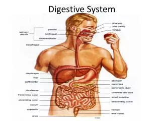
DIGESTIVE SYSTEM. ORAL CAVITY. Oral cavity consists of the mouth and its structures, which include the tongue, teeth and their supporting structures ( periodontium ), major and minor salivary glands, and tonsils. Divided into vestibule and oral cavity proper . MUCOSA.

E N D
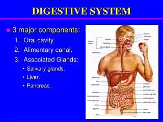
ORAL CAVITY • Oral cavity consists of the mouth and its structures, which include the tongue, teeth and their supporting structures (periodontium), major and minor salivary glands, and tonsils. • Divided into vestibule and oral cavity proper.
MUCOSA • A mucous tissue lining various tubular structures consisting of epithelium, lamina propria, and, in the digestive tract, a layer of smooth muscle (muscularismucosae).
MUCOSA OF ORAL CAVITY Masticatory mucosa • Has a keratinized and parakeratinized stratified squamous epithelium • Found in gums and hard palate
Lining mucosa Non keratinized epithelium Few papillae Found on lips, cheeks,floor of oral cavity
Specialized mucosa Associated with taste sensation Restricted to dorsal surface of tongue
LIPS • Cutaneous area • Red area • Oral mucosa
CHEEKS • Skin with sebaceous and sweat glands • Mucosa lined by Stratified squamous non cornified epithelium • Lamina propria is compact, contain papilla and connected by a submucosa to the underlying skeletal muscle. • Submucosa has elastic fibers and tubuloalveolar glands
GUMS • Epithelium • Lamina propria • No submucosa and no glands
HARD PALATE • Epithelium keratinized • Long vascular papillae • Periosteum of the hard palate • Submucosa with collagen fibers and gland
SOFT PALATE • Oral surface • Pharyngeal surface • Mucosa • Submucosa • Glands • Skeletal muscles
TONGUE • The main bulk of the tongue, particularly of the anterior two-thirds, is skeletal muscle.
The interlacing muscle fibers course chiefly in three directions, longitudinally, transversely, and vertically, an arrangement which gives maximal mobility and physical control.
LOWER SURFACE • Stratified squamousnoncornified epithelium • Lamina propria is thin and closely bound to underlying muscle.
DORSAL SURFACE • Anterior 2/3: Lingual papillae • Posterior 1/3: Mucosal ridges and lingual tonsils • No submucosa
FILIFORM PAPILLAE • Most numerous • Smallest • Evenly distributed • Slender core of vascular epithelium • Stratified squamous epithelium • Secondary projections
FUNGIFORM PAPILLAE • Few in number • Interspersed among the filiform papillae • Rounded summits and broader bases • Noncornified epithelium • Highly vascular connective tissue
1.STRATIFIED SQUAMOUS NONCORNIFIED EPITHELIUM 2.LAMINA PROPRIA 3.TASTE BUD
FOLIATE PAPILLAE • Along the posterolateral border of the tongue there are folds of the mucous membrane, sometimes called the foliate papillae. • Not well developed in humans. • In some animals,e.g.,rabbits, it constitute the principle site of aggregation of taste buds.
CIRCUMVALLATE PAPILLAE • 9-12 in number • Resemble fungiform papillae • Much larger • Surrounded by a trench and a wall • Small oval bodies, taste buds
DORSAL SURFACE OF THE POSTERIOR THIRD OF THE TONGUE • Free of papillae • Has mucosal ridges and lingual tonsils
ANATOMICAL DIVISION • Crown • Root • Neck
COMPONENTS OF TOOTH • Enamel • Dentin • Cementum • Pulp
ENAMEL • Consists of Enamel rods Inter rod enamel • Secreted by ameloblasts • At apical pole------Tomes process • At the base---------cluster of mitochondria
ENAMEL • Acellular mineralized tissue that covers the crown of tooth • Hardest substance in the body • Once formed, can't be replaced • 96-98% exist mainly as hydroxyapatite crystals
DENTIN • Calcified tissue harder than bone • Dentin is produced by neural crest derived odontoblasts of the adjacent mesenchyme • Sensitive structure
Cementum Alveolar bone proper Periodontal ligament Gingiva PERIODONTIUM
PULP • A C.T. compartment bounded by the tooth dentin • Apical foramen • Vascular & neural networks • Cavity decreases with age