Download
1 / 38
380 likes | 459 Views
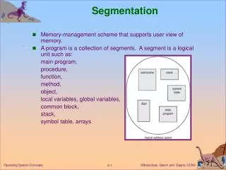
Segmentation Foundations. Easy Segmentation Tissue/Air (except bone in MR) Bone in CT Feasible Segmentation White Matter/Gray Matter: MRI M.S. White Matter Lesions: MRI. Statistical Classification. Probabilistic model of intensity as a function of (tissue) class Intensity data

E N D
Segmentation Foundations • Easy Segmentation • Tissue/Air (except bone in MR) • Bone in CT • Feasible Segmentation • White Matter/Gray Matter: MRI • M.S. White Matter Lesions: MRI
Statistical Classification • Probabilistic model of intensity as a function of (tissue) class • Intensity data • Prior model Classification of voxels [Duda, Hart 78][MRI: MikeVannier late 80s]
p(x|tissue class J) intensity probability density mean for tissue J Measurement Model • Characterize sensor Tissue class conditional model of signal intensity
A bit of notation… • Estimate by finding the one that maximizes the function f
Maximum Likelihood (ML) Estimation • Estimate parameters to maximize probability of observed data conditioned on parameters . • yo : observed data • p(y|) : Measurement Model • : Model Parameters
p(x|gray matter) p(x|white matter) intensity Example
gray matter white matter threshold Example - revisited
Multiple Sclerosis T2w PDw Provided by S Warfield
Dual Echo MRI Feature Space csf T2 Intensity severe lesions air wm gm PD Intensity
severe csf lesions mild healthy gm wm Detail • MS Lesions are “graded phenomenon” in MRI, and can be anywhere on the curve
Multiple Sclerosis T2w Segmentation PDw Provided by S Warfield
Maximum A-Posteriori (MAP) Estimation • Estimate parameters to maximize posterior probability model parameters conditioned on observed data • Use Baye’s rule – ignore denominator • p() : Prior Model
Provided by S Warfield Multiple Sclerosis PDw T2w kNN SVC
Background: Intensity Inhomogeneities in MRI • MRI signal derived from RF signals… • Intra Scan Inhomogeneities • “Shading” … from coil imperfections • interaction with tissue? • Inter Scan Inhomogeneities • Auto Tune • Equipment Upgrades
ML Estimation – with missing data • x : missing data (true labeling) • y0 : observed intensities • : (parameters of) bias field
ML Estimation – EM Approach • E []: Expected value under p(x|yo, ) • Take expectation of objective function with respect to the missing data, conditioned on everything we know • x : missing data (true labeling) • y0 : observed intensities • : (parameters of) bias field
EM Algorithm • General exponential family • Iterate to convergence: M step: E step:
EM Algorithm: Example • Measurement Model • Tissue intensity properties with bias correction • Missing Data • Unknown true classification • Prior Models • Tissue Frequencies • Intensity Correction is Low Frequency • ML estimate of bias
EM-Segmentation E-Step Compute tissue posteriors using current intensity correction. Estimate intensity correction using residuals based on current posteriors. M-Step Provided by T Kapur
EM Segmentation… Seg Result w/o EM Seg Result With EM PD, T2 Data
EM Segmentation… External Surface of Brain
EM Segmentation… WM Surface with EM WM Surface w/o EM
EM Segmentation: MS Example PD T2 Data provided by Charles Guttmann
EM Segmentation: MS Example Seg w/o EM Seg with EM
Prior Probability Models • Simple: Frequency of Tissues • More Interesting: • Powerful Mechanism for Incorporating Domain Knowledge into Segmentation • Tissue properties • Relative Location of Structures • Atlases
Prior Model Example: EM-MF Segmentation • Tina Kapur PhD thesis • EM Segmentation, augmented with • Ising prior of tissue homogeneity • Solved with Mean Field Approxomation • Prior on relative position of organs • Spatially Conditioned Models
Prior Models: Ising Model • Ising Model can capture the phenomenon of piecewise-homogeneity. • Initially used in Statistical Physics to model the magnetic domains in Ferromagnetism. • Used in Medical Image Processing to model the piecewise-homogeneity of Tissue.
Prior Models: Ising Model • Ising Model relaxes spatial independence assumption • Voxels depend conditionally on (only) their neighbors • More probable to agree with neighbor
Define the Neighborhood 1st Order Lattice 2nd Order Lattice 6 Neighbors 26 Neighbors Reduce calculation cost => use 1st order Lattice Neighbors = {East, South, West, North, Up, Down} Provided by K Pohl
Potts Model • Potts model generalizes Ising model so that each lattice site takes on several values (more than two). • Frequently used to model tissues (e.g. White Matter, Gray Matter, CSF, Fat, Air, etc.)
Some Results EM EM-MF Provided by T Kapur
More Results Noisy MRI EM Segmentation EM-MF Segmentation Provided by T Kapur
Posterior Probabilities (EM) White matter Gray matter Provided by T Kapur
Posterior Probabilities (EM-MF) White matter Gray matter Provided by T Kapur
Segmentation of 31 Structures Kilian Pohl PhD (defense several weeks ago)
Segmentation of 31 Structures Lower Front Provided by Kilian Pohl
Segmentation of 31 Structures Superior Temporal Gyrus Provided by Kilian Pohl