Download
1 / 57
590 likes | 768 Views
Adrenals. Objectives:. History. Embryology. Anatomy. Physiology. Imaging. Surgical Diseases: Incidentaloma Conn’s Syndrome. Pheochromocytoma/ Paraganglioma . Cushing disease VS Cushing Syndrome. Adrenocortical carcinoma . Operative approaches . History. 1563 anatomy

E N D
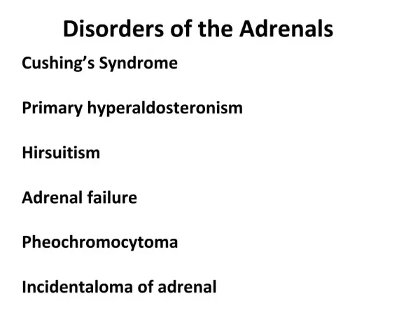
Objectives: • History. • Embryology. • Anatomy. • Physiology. • Imaging. • Surgical Diseases: • Incidentaloma • Conn’s Syndrome. • Pheochromocytoma/ Paraganglioma. • Cushing disease VS Cushing Syndrome. • Adrenocortical carcinoma. • Operative approaches.
History • 1563 anatomy • 1855 Addison described clinical features of the syndrome named after him. • 1912 Cushing described hypercortisolism. • 1934 the role of adrenal tumors in hypercortisolism understood. • 1955 pheochromocytoma was first described by frankel.
Embryology • Paired gland • Cortex (coelomic epithelium). • Zona glomerulus Mineralocorticoid • Zona fasciculateGlucocorticoid • Zona reticularis.(3rd year)Sex hormones • Medulla( ectoderm) neural crest. • Ectopic tissue.
Physiology: • Adrenal cortex • Aldosterone • Cortisol • Sex steroids • Adrenal medulla: • Noradrenaline (20%). • Adrenaline (80%).
Adrenal Imaging: • CT scan: • Benign • Intensity similar to liver • Low attenuation • Homogeneous • Smooth border • Smooth contour • < 4 cm in greatest dimension
Cont CT scan • Malignant lesions: • High attenuation (>30 HU). • Heterogeneous. • Irregular borders. • Local/ vascular invasion. • Lymphadenopathy. • Metastases. • Large size (>6cm).
Radiology: • MRI. • Nuclear scan. • PET scan.
Incidentaloma • Found in 1-4 % of CT scans. • Increases with age. • Small nonfunctioning adrenal tumors. • some with subclinical secretions of hormones. • Adrenocortical carcinoma. • Metastases.
Incidentaloma: • Nonfunctioning adenoma 82% • Subclinical Cushing 5% • Pheochromocytoma 5% • Adrenocortical ca 5% • Metastatic carcinoma 2% • Conn’s 1%
Diseases of The Adrenals • 1- hyperaldosteronism • Causes: • Primary • Adenoma. • Idiopathic bilateral adrenal hyperplasia. • Unilateral adrenal hyperplasia. • Adrenocortical carcinoma. • Familial • Secondary • Renal artery stenosis. • CHF. • Liver cirrhosis. • Pregnancy.
Primary hyperaldosteronism: • Age 30-50 years • Female> male, 2:1 • Prevalence 5-13% • HPT with or without hypokalemia. • Weakness, polyuria, paresthesis, tetany, cramps. • Metabolic alkalosis, relative hypernatremia. • Elevated aldosterone secretion and suppressed plasma renin activity.
Cont: • Screening tests: • PAC (ng/dl) / PRA (ng /ml)>20. • Plasma aldosterone >15 ng/dl. • Confirmatory tests: • Sodium suppression test • Urinary aldosterone excretion >14 ug/ 24hr.
Treatment: • Pre-operative preparation: • Spironolactone: Competitive aldosterone antagonist • Promote K retention. • Reduce extracellular volume . • Reactivate the renin-angio-aldosterone syst. • Amiloride: K sparing diuretics
Cont. • Surgery: • Laparoscopic adrenalectomy. • Open surgery. • Medical treatment: • Unfit patients. • Bilateral ald.
Prognosis: • 1/3 persistent hypertension. • K level will be restored.
Pheochromocytoma: EPIDEMIOLOGY: • Less than < 0.1% of patients with hypertension • 5% of tumors discovered incidentally on CT scan • Most occur sporadically • •Associated with familial syndromes, such as: _Multiple endocrine neoplasia type 2A (MEN 2A) • –MEN 2B
Cont. –Recklinghausen disease –von Hippel-Lindau disease • Pheochromocytomas are present in 40% of patients with MEN 2 • 90% of patients with pheochromocytoma are hypertensive • • Hypertension less common in children • • In children, 50% of patients have multiple or extra-adrenal tumors
Symptoms and signs: • Clinical findings are variable • Episodic or sustained hypertension • Triad of palpitation, headache, and diaphoresis • Anxiety, tremors and Weight loss. • Dizziness, nausea, and vomiting • Abdominal discomfort, constipation, diarrhea.
Cont: • • Visual blurring • • Tachycardia, postural hypotension • • Hypertensive retinopathy
Cont: ESSENTIAL FEATURES • Episodic headache, excessive sweating, palpitations, and visual blurring • Hypertension, frequently sustained, with or without paroxysms • Postural tachycardia and hypotension • • Elevated urinary catecholamines or their metabolites, hyper metabolism, hyperglycemia
Cont. • • Rule of 10s: • – 10% malignant • –10% familial • –10% bilateral • –10% multiple tumors • –10% extra-adrenal
Cont. Extra-adrenal pheochromocytomas: –Abdomen (75%) –Bladder (10%) –Chest (10%) –Pelvis (2%) –Head and neck (3%)
LABORATORY FINDINGS • Hyperglycemia • Elevated plasma metanephrines • Elevated 24-hour urine metanephrines and free catecholamines • Elevated urinary vanillylmandelic acid (VMA) • Elevated plasma catecholamines
IMAGING FINDINGS • Adrenal mass seen on CT or MRI • Characteristic bright appearance on T2-weighted MRI • Asymmetric uptake on MIBG scan. Particularly useful for extra-adrenal, multiple, or malignant pheochromocytomas. • MIBG Not useful for sporadic biochemical syndrome with unilateral mass
DIAGNOSTIC CONSIDERATIONS: • Avoid arteriography or fine-needle aspiration as they can precipitate a hypertensive crisis • Early recognition during pregnancy is key because if left untreated, half of fetuses and nearly half of the mothers will die
RULE OUT: • Other causes of hypertension • Hyperthyroidism • Anxiety disorder • Carcinoid syndrome
WORK-UP: • History and physical exam • Suspect pheochromocytoma based on symptoms • CT, MRI, or other scans • Plasma and urine studies (metanephrines, catecholamines, VMA) • Begin treatment with a-blockers • Possible MIBG scan • • Operative excision of tumor
WHEN TO admit: • Hypertensive crisis (can develop multisystem organ failure, mimicking severe sepsis
TREATMENT AND MANAGEMENT: • a-Adrenergic blocking agents should be started as soon as the biochemical diagnosis is established to restore blood volume, to prevent a severe crisis, and to allow recovery from the cardiomyopathy • SURGERY: • Indications: • • All pheochromocytoma should be excised • Contraindications: • • Metastatic disease • • Inadequate medical preparation (a- blockade)
Cushing disease VS Syndrome • Cushing disease secondary to pituitary adenoma. • Cushing syndrome secondary to anything else.
Adrenocortical carcinomafunctioning VS non functioning: ESSENTIAL FEATURES : • Variety of clinical symptoms through excess production of adrenal hormones • Complete surgical removal of the primary lesion and any respectable metastatic sites has been the mainstay of treatment
EPIDEMIOLOGY: • These tumors are rare; 1—2 cases per million persons in the United States • Less than 0.05% of newly diagnosed cancers per year • Bimodal occurrence, with tumors developing in children < 5 years of age and in adults in their fifth through seventh decade of life • • Male:female ratio is 2:1, with functional tumors being more common in women
Cont: • • Left adrenal involved slightly more often than the right (53% vs 47%); bilateral tumors are rare (2%) • • 50—60% of patients have symptoms related to hypersecretion of hormones (most commonly Cushing syndrome and virilization) • • Feminizing and purely aldosterone-secreting carcinomas are rare • • 50% of patients have metastases at the time of diagnosis
SYMPTOMS AND SIGNS: • Symptoms of specific hormone excess (cortisol excess, virilization, feminization) • Palpable abdominal mass • Abdominal pain • Fatigue, weight loss, fever, hematuria
LABORATORY FINDINGS: • All laboratory abnormalities depend on hormonal status of tumor • Elevated urinary free cortisol or steroid precursors • Loss of normal circadian rhythm for serum cortisol • Low serum adrenocorticotropic hormone (ACTH) • Abnormal dexamethasone suppression test • Elevated serum testosterone, estradiol, or aldosterone levels
IMAGING FINDINGS: • Evaluation of adrenal glands with CT or MRI (adrenocortical carcinomas are typically isodense to liver on T1-weighted MRI, and hyperdense relative to liver on T2-weighted MRI images) • MRI more accurately gauges the extent of any intracaval tumor thrombus
DIAGNOSTIC CONSIDERATIONS: • Mean diameter of adrenal carcinoma at diagnosis is 12 cm • Radiographic evaluation of suspected metastatic sites for purposes of staging should be undertaken prior to thought of any surgery • RULE OUT Pheochromocytoma