Download
1 / 30
330 likes | 421 Views
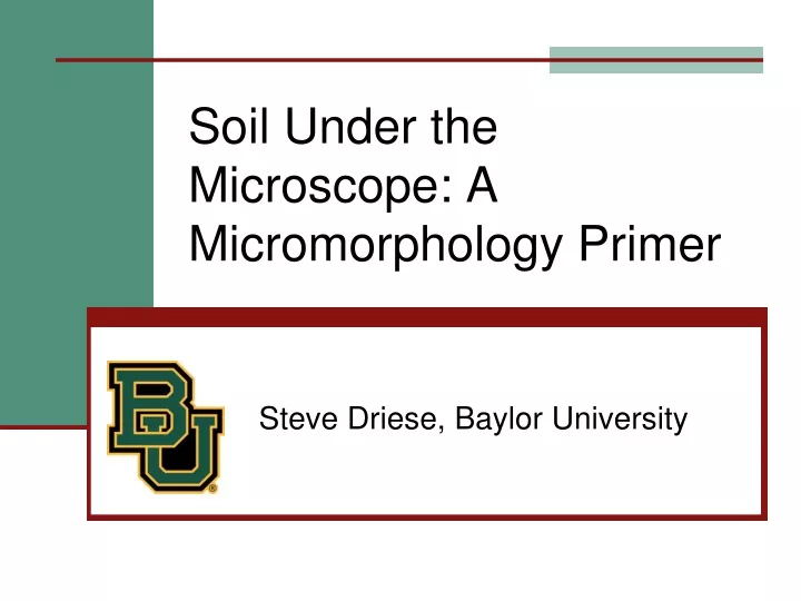
Soil Under the Microscope: A Micromorphology Primer. Steve Driese, Baylor University. Introduction. What is soil “micromorphology”? Why is it useful for interpreting genesis of soils? Integrate in undergraduate sedimentary petrology class, or as part of a soils course?

E N D
Soil Under the Microscope: A Micromorphology Primer Steve Driese, Baylor University
Introduction • What is soil “micromorphology”? • Why is it useful for interpreting genesis of soils? • Integrate in undergraduate sedimentary petrology class, or as part of a soils course? • Can be applied interpreting soil drainage, soil maturity, hydrogeology, and contaminant fate and transport.
What is “Micromorphology”? • Application of basic geologic thin-section petrography to the study of soils and paleosols • Terminology differences can seem daunting! Pledger series Vertisol on 6,000 yr-old floodplain surface, Dance Bayou, Brazoria County, TX
You may feel like it’s too much to swallow!.......But just hang in there.
Basic Terminology At the very basic level soil consists of: • Soilmatrix, termed “S-matrix” by soil scientists. • Pedologicalfeatures with origins related to soil-forming processes.
From Brewer (1976): Soil Matrix • Plasma, mainly fine clay-sized mineral particles, but also including organic material of colloid size, which may be soluble, • Skeletongrains, chiefly silicate sand and silt grains embedded in the plasma, which are generally stable, and • Soilvoids, which are pore spaces occupied by air or water, and include: (A) Macropores (>1-2 µm) such as root pores, animal burrows, interpedal and fracture pores, and (B) Micropores (< 1-2 µm).
Pedological Features Inherited Features: lithorelicts (pieces of parent material), saprorelicts (saprolitized parent material) and pedorelicts (reworked soils). 1.5 mm, PPL 5.5 mm, XPL
Pedological Features 2. Features due to plasma concentrations: nodules (non-banded and layered) and concretions (banded and layered), 1.5 mm, XPL 1.5 mm, XPL
Pedological Features 3. Features due to plasma separations: cutans (coatings in voids or on grain surfaces), sepic-plasmic fabrics (bright- or flecked clays) and crystic plasmic fabrics (crystals showing displacive growth), XPL XPL 1.5 mm, PPL
Pedological Features 3. Features due to plasma separations: cutans (coatings in voids or on grain surfaces), sepic-plasmic fabrics (bright- or flecked clays) and crystic plasmic fabrics (crystals showing displacive growth), XPL XPL
Pedological Features 3. Features due to plasma separations: cutans (coatings in voids or on grain surfaces), sepic-plasmicfabrics (bright- or flecked clays) and crystic plasmic fabrics (crystals showing displacive growth), 1.5 mm, XPL 0.5 mm, XPL
Pedological Features 3. Features due to plasma separations: cutans (coatings in voids or on grain surfaces), sepic-plasmic fabrics (bright- or flecked clays) and crysticplasmicfabrics (crystals showing displacive growth), 0.5 mm, XPL 1.5 mm, XPL 1.5 mm, XPL
Pedological Features 4. Features due to biological activity: (biotic tubules filled with various kinds of soil materials, including fecal materials), PPL UV Fluorescence
Pedological Features 4. Features due to biological activity: (biotic tubules filled with various kinds of soil materials, including fecal materials), 1.5 mm, XPL
Pedological Features 4. Features due to biological activity: (biotic tubules filled with various kinds of soil materials, including fecal materials), 1.5 mm, PPL 1.5 mm, PPL
Pedological Features: Paleosol Examples – Illuviated clays XPL
Pedological Features: Paleosol Examples– Carbonized roots XPL
Pedological Features: Paleosol Examples – Fe-depletion root PPL
Pedological Features: Paleosol Examples – Rhizolith + Fe depletion root trace Calcite rhizolith around silt- and clay- infilled root trace 7 cm, PPL Redox-depletion and enrichment quasi-coatings with root trace 5.5 mm, PPL
Pedological Features: Paleosol Examples – Sepic-plasmic fabrics 0.5 mm, XPL
Pedological Features: Paleosol Examples – Fe-Mn concretions 1.5 mm, PPL 1.5 mm, XPL
Pedological Features: Paleosol Examples – Calcite nodule XPL
Pedological Features: Paleosol Examples – Meniscate burrow PPL
Pedological Features: Paleosol Examples – Thecate amoebae UV Fluorescence
A Hierarchical Approach to Thin-Section Description (after Bullock et al., 1985)
Micromorphology • Useful, for evaluating soil mineralogy, genesis, and diagenetic processes (Cady et al., 1986). • Inexpensive, simple: the cost of a thin-section and a petrographic microscope. • Essential precursor for followup analytical procedures (e.g., geochemistry, isotopes).
References Cited Brewer, R., 1976, Fabric and Mineral Analysis of Soils, 2nd edition: Huntington, New York, Robert E. Krieger Publishing Co., 482 p. Bullock, P, Fédoroff, N., Jungerius, A., Stoops, G., Tursina, T., and Babel, U., 1985, Handbook for Soil Thin Section Description: Wolverhampton, UK, Waine Research Publications, 152 p. Cady, J.G., Wilding, L.P., and Drees, L.R., 1986, Petrographic microscope techniques, in Methods of Soil Analysis, Part I. Physical and Mineralogical Methods: Soil Science Society of America, Monograph No. 9, p. 185-218. Stoops, G., 2003, Guidelines for Analysis and Description of Soil and Regolith Thin Sections: Madison, WI, Soil Science Society of America, 184 p. + CD w/images. Stoops, G., Marcelino, V., and Mees, F. (eds.), 2010, Interpretation of Micromorphological Features of Soils and Regoliths: Amsterdam, Elsevier Pub. Co., 720 p.