Download
1 / 126
1.66k likes | 3.48k Views
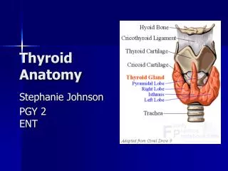
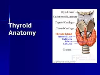
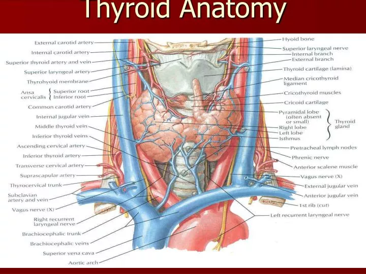
Thyroid Anatomy. Brownish-red, highly vascular gland Location: ant neck at C5-T1, overlays 2 nd – 4 th tracheal rings Avg. width: 12-15 mm (each lobe) Avg. height: 50-60 mm long Avg. weight: 25-30 g in adults (slightly more in women) **enlarges during menstruation and pregnancy**

E N D
Brownish-red, highly vascular gland • Location: ant neck at C5-T1, overlays 2nd – 4th tracheal rings • Avg. width: 12-15 mm (each lobe) • Avg. height: 50-60 mm long • Avg. weight: 25-30 g in adults (slightly more in women) **enlarges during menstruation and pregnancy** Pyramidal lobe: • often ascends from the isthmus or the adjacent part of either lobe (usu L) up to the hyoid bone • may be attached by a fibrous/fibromuscular band “levator” of the thyroid gland
Transverse view: relationship to other NB structures in neck
Relation w/ Strap muscles • Lateral – sternothyroid • Anterior -omohyoid muscle • sternohyoid • Inferior - SCM (lower portion) ** careful - motor nerve supply from the ansacervicalis enters these muscles inferiorly.
Vascular Anatomy ARTERIAL: • superior and inferior thyroid arteries (occthyroideaima) • ++ collateral anastomoses (ipsi and contralaterally) • thyroid ima (when pres) originates from aortic arch or innominate artery, enters the thyroid at inferior border of isthmus.
Vascular anatomy VENOUS: 3 pairs of veins: • STV – asc along STA and becomes a tributary of the IJV • MTV – directly lateral IJV • ITV (variable): • R – passes ant to innominate a R BCV or ant trachea L BCV • L – drainage L BCV **occ – both inf veins form a common trunk “thyroid ima vein” empties into L BCV
Relationship with RLN: • RLN ascends in the TE groove and enters the larynx b/w the inferior cornu of the thyroid cartilage and the arch of the cricoid • RLN can be found after it emerges from the superior thoracic outlet: • Sup: thyroid lobe • Lat: common carotid artery • Medial: trachea
Lymphatics • Extensive, multidirectional flow • periglandular prelaryngeal (Delphian) pretracheal paratracheal (along RLN) brachiocephalic (sup mediastinum) deep cervical thoracic duct
Structure • Under pretracheal thyroid inner true capsule thin and closely adherent to the gland • capsule extensions within the gland form septae, dividing it into lobes and lobules • lobules are composed of follicles = structural units of the gland layer epithelium enclosing a colloid-filled cavity • colloid (pink on H&E stain) contains an iodinated glycoprotein, iodothyroglobulin (precursor of thyroid hormones).
Structure Epithelial cells = 2 types: • principal (ie: follicular) – formation of colloid (iodothyroglobulin) • parafollicular (ie: C cells -clear, light), lie adjacent to follicles w/in basal lamina produce calcitonin
THYROID GLAND HISTOLOGY http://arbl.cvmbs.colostate.edu/hbooks/pathphys/endocrine/thyroid/anatomy.html
Thyroid Hormone Synthesis • 1. Iodide trapping • 2. Oxidation of iodide and iodination of thyroglobulin • 3. Coupling of iodotyrosine molecules within thyroglobulin (formation of T3 and T4) • 4. Proteolysis of thyroglobulin • 5. Deiodination of iodotyrosines • 6. Intrathyroidal deiodination of T4 to T3
OH OH I I I I I O O NH2 NH2 I I OH OH O O 3,5,3’-Triiodothyronine (T3) THYROID HORMONES Thyroxine (T4)
THYROID HORMONES IN THE BLOOD • Approximately 99.98% of T4 is bound to 3 serum proteins: Thyroid binding globulin (TBG) ~75%; Thyroid binding prealbumin (TBPA or transthyretin) 15-20%; albumin ~5-10% • Only ~0.02% of the total T4 in blood is unbound or free. • Only ~0.4% of total T3 in blood is free.
NH2 NH2 I OH I OH OH “Step up” “Step down” I O O O I T4 I I OH I R OH I R I O O I T3 rT3 I OH I R O 3,3’-T2 R = THYROID HORMONE METABOLISM
Effects of Thyroid Hormone • Fetal brain and skeletal maturation • Increase in basal metabolic rate • Inotropic and chronotropic effects on heart • Increases sensitivity to catecholamines • Stimulates gut motility • Increase bone turnover • Increase in serum glucose, decrease in serum cholesterol • Increases oxygen consumption in most target tissues.
Disorders of Thyroid: • Functional-Hyperthyroidism • Hypothyroidism • (Euthyroid) • Thyroiditis. • Neoplasms – adenoma/carcinoma. • Congenital – Thyroglossal cyst/duct.
History • Period-Duration.Days.Weeks.Months.years • Progress-Rapid, slowly • Pressure symtopms.Dyspnea.Dysphagia • Palpitation.TG • Pain-Thyroiditis • Paralysis-Change of voice.Malignancy?
Pertinent questions –clinical assessment of goiter • 1-is it Goiter-? • 2-Diffuse or Nodular? • 3-Single nodule –Solitary thyroid nodule or Dominant nodule in MNG 4- Function wise: Hyper, Hypo or euthyroid? 5-Thyroiditis? 6-Signs of malignancy? 7-Retrosternal Extension? 8-recurrent
EXAMPLES OF THYROID DISEASES 1° Hypothyroidism Hyperthyroidism Congenital Hypothyroidism www.hsc.missouri.edu/~daveg/thyroid/thy_dis.html
Thyroid Disease Spectrum Overt Hypothyroidism TSH >4.0 IU/mL, Free T4 Low Mild Thyroid Failure TSH >4.0 IU/mL, Free T4Normal Euthyroid TSH 0.4-4.0 IU/mL, Free T4 Normal Thyrotoxicosis TSH <0.4 IU/mL, Free T3/T4Normal or Elevated 10 0 5 TSH, IU/mL Braverman LE, et al. Werner & Ingbar’s The Thyroid. A Fundamental and Clinical Text. 8th ed. 2000. Canaris GJ, et al. Arch Intern Med. 2000;160:526-534. Vanderpump MP, et al. Clin Endocrinol (Oxf). 1995;43:55-68.
Classification of goiter1-Simple nontoxic a-Diffuse hyperplastic-i-physiological-puberty.preg.ii-prim.Iodine def-Endemic G iii-Sec.Iodine def. *Goiterogens-of Brassica family-Cabbage,soya bean *Excess dietary flouride. *Drugs-PAS,Lithium,Phenylbutazone. Thiocyanates,potassium perchlorate. Antithyroiddrugs.radioactiveiodine.Dyshormonogenesis b-Colloid G c-MNG d-Solitary nontoxic nodule e-Recurrent nontoxic nodule
2-Toxic Ga-Diffuse-prim- Graves’ disb-MN(sec.)Plummer’s disc-Toxic nodule-Solitary-Tertiary-Toxic adenomad0 Recurrent toxic G3-Thyroiditisa-acuteb-subacutec-chronic-CLT4-neoplastic a.benignb-malign
Solitary thyroid nodule(STN)Aims-1-Determine whether it is causing localized or systemic symptoms2-Whether it is benign or malignantDDX:1-Benign G2-Cyst(intrathyroidal)3-thyroiditis4-Benign tumor5-Malignant tumorHy- IND- Sex-STN-more likely to be Ca in a man than woman Age= = = in young (<20) & older >60Y Thyroid Ca occurs in 40% of children withSTN Residence(place of birth)-Benign nodule in endemic G areas 6 Ps Past Hy-Hyvof radiation to neck( most important)-Low –dose therapeutic radiation(6.5-2000cGy) in infancy & childhood—Increased incidence of benign G(35%), or thyroid cancer(13%) family Hy-Thyroid Ca is familial in 25% of patientswith Medullary Ca O/E- Palpate systematically – STN or MNG Solitary hard nodule-likely to be mailgnant Cervical LAP-?Investigations:US– Solid or cystic ? number of nodules Suspicious nodule? Coexistent suspicious LN?FNA + US guideCytological results: 1-Mailgnant 2-Benign 3-Suspicious 4-Inadequate repeat biopsy False +ive is rare 20% of suspicious- & 5% of benign reports-are actually malignant CXR—including neck-Tracheal displacement.calcification of nodule.Pul.metastasisIndications for surgery_1-suspicion or documented Ca.2-Pressure symptoms.3-TG4-Substernal extension5-Cosmetic deformityNon-op.Rx-1-small or moderately sized MNG2-CLT.unless suspicious area.3-Hy of radiation-4-Family Hy of Ca. thyroid
MNGPathogenesis:1-Persistent TSH stimulation---Diffuse hyperplasia of gland( all active lobules)---later with 2-fluctuation of TSH level-----mixed areas of active & inactive lobules develop-----3-active lobules become more vascular & hyperplastic4-Hage occur with necrosis in the center5- nodule formation6- center of nodule is inactive& only margin is active i.einternodular tissue is active----7- Formation of many nodules---MNG
Colloid G-Due to long standing Iodine deficency + localized accumulation of significant colloid in the glandMNG: Clinical features-1-More common in middle aged females2-long Hy-many years. slowly progressive3-Many nodules. Dif.size, in both lobes , isthmus4- nodule- firm, nontender & moves with deglutition.5-Recent increase in size-short duration- hageweeks to months- malign transformation
Complications of MNG1-Sec thyrotoxicosis2-Follicular Ca. of thyroid3-Hage in a nodule4-tracheal compression- -retrosternal extension/obstruction5-Cosmetic problemInvestigationsTFTs-TSH, F(T4)U/SFNAXR of neck- calcification. Position & compression of trachea.Rx Surgery S/TNear total thyroidectomyTotal T
Solitary thyroid noduleCauses1-Thyroid cyst2-Thyoid adenomasa- Follicularb-Hurthle cell3- Papillary Ca4-Dominanent nodule in MNGTypes:1- Toxic adenoma2-Nontoxic Solitary nodulebased on radioactive study:a- Hot –autonomous toxic noduleb-Warm-normally functioning nodulec-Cold-nonfunctioning-thyroiditis, thyroid cyst, hage, malig
Clinical features1-Single palpable nodule2- progress- rapid enlargement may be malig3-Age – extreme of life – can be malig.4-30 % are cystic4- up to 20 % of cold nodule –malig5-site- commonest site at junction of isthmus wit one of lat lobe.Clinical features suggesting malig:1-any nodule can be malig2-rapid onset/or recent increase in size.3-Pressure effects4-paralysis- hoarseness of voice5-hard, irregular, fixed nodule6-palpable significant cervical LAP
Investigations1-TFTs2-neck U/S3-FNA4-Radioactive isotope study(Iodine 123,131, Tc 995-XR- neck- tracheal deviation.Rxindications for surgery:1-Cyst->4cm.Hagic,Malig or suspecios on FNA, Recurrent Cyst. Complex cyst (both solid & cystic component).2-follicular neoplasm3-malig nodule4-toxic adenoma in young patient5- pressure effects6- Cosmetic reason
Rx-continuedA- Conservative- Colloid nodule- T4 Rx- respond in 50%if nodule: reappears,or enlarges rapidly or causes cosmetic problem—then -HT(hemithyroidectomyB-Surgery- minimal op.HT1-Nontoxic G- hemithyroidectomy including the isthmus.2-Papillary Ca. near total T.+suppression dose of T4(0.2-0.3mg/d)3-Toxic adenoma-age >45; radioactive iodine(131) 5 milli curie orally.Age <45Contro with antithyroid then surgery- hemithyrodectomy4-If FNA-follicular adenoma-hemithyroidectomyIf HPE- follicular Ca(capsular & vascular invasion) then complete total T( W/N1wk or after 3wks)5-if nodule in the isthmus- isthmusectomy + part of adjacent lat. lobes6-If FNA- Medullary Ca-Total T+bilat.neck nodal dissection including central compartment
Thyroid cyst1- Tensly cystic swelling may be hard-2- common cause – colloid degeneration. 50%- absence of epithelial lining3- 30% of solitary nodules are cystic4- 15% cystic swellings- are malig5-cystic formation is common in papillary ca.6-complex cyst- contains both cystic & solid areas , more likely to be mlig7- FNAC may cause regression of simple cyst.8- Recurrence after 3 aspiration or if cyst hagic –surgery9-Complex cyst or > 4cm or malig or suspecious- surgery
SOB in Goiter1-MNG- tracheal compression2- Retosternal G3- Sec toxic G- CHF4- Ca. infiltrating the trachea
Thyrotoxicosis & HyperthyroidismThyrotoxicosis- symptoms complex caused by increased levels of thyroid hormones’Types:1-Diffuse toxic G- graves’ dis- Prim thyrotoxicosis.2-toxic MNG-Sec Thyrotoxicosis3- Toxic adenoma- nodule4- Others:a-thyrotoxicosisfactitia- drug induced . Excessive intake of T4 b-JodBasedowthyrotoxicosis- by large doses of iodine given to hyperplastic endemic Gc-Thyroiditis- de Quervain’s or Autoimmuned-Occ. Ca thyroide-Neonatal thyrotoxicosis- subsides in 3-4 wksf- Struma ovariig- Drugs-amiodarone- antiarrhythmic agent
Clinical featuresIncidenceAge- anySex- F: M 8:1Prim. Commonly in younger age groupSec. is common in older gae groupGraves’ dis- may present without obvious goiterSuspect Graves’ disese In:1-Unexplained behavioural problem2-insomnia3-myopathy4-Unexplained diarrhea or wt loss5-Tachycardia6-menstrual changes or infertility
Symptoms1-CNS-Irritability.Nervousness. Insomnia2-Neuromascular system:Undue fatigue & muscle weakness.Tremor3-Skeletal system: increase in linear growth in children4-Skin; hair loss . Pruritus.palmarerythema.5-CVS:tachycardai.SOB at rest or on mild exersion.Angina. Arrythmias.CHF(in elderly)6-GIT-Wt loss despite good appetite.diarrhea( due to increased activity at ganglionic level).7-GUT: oligo or amenorrhea.Occ. Urinary frequency
Sympathetic overactivity:causes-SOB,Palpitation.tiredness.Heatintolerance.Sweating,nervousness,increased appetite & decrease in Wt.Because of increased catabolism they have increased appetite , decrease wt & also increased creatinine level which signifies myopathy due to more muscle catabolism.Fine tremor_ due to diffuse irritability of gray matter.Thrill & bruit are detected in upper pole as the Sup TA enters the gland superficially while ITA enters from deeper plane so the thrill cannot be felt in lower pole
THYROID EYE DISEASE • INFILTRATION • 1. soft tissue involvement :- chemosis, conjunctival injection over the recti insertions, puffy lids
THYROID EYE DISEASE • Superior limbic keratoconjunctivitis (SLK)
Clinical Characteristics of Exophthalmos • Proptosis • Corneal Damage • Periorbital edema • Chemosis • Conjunctival injection • Extraocular muscle impairment • Optic neuropathy
Clinical Characteristics of Localized Myxedema • Raised surface • Thick, leathery consistency • Nodularity, sometimes • Sharply demarcated margins • Prominent hair follicles • Usually over pretibial area • Non-tender
Margins sharply demarcated Margins sharply demarcated Graves’ Disease - Localized Myxedema Nodularity Thickened skin