Download

1 / 23
230 likes | 331 Views
Anesthetic Methods in the Management of Carotid Endarterectomies. Daniel Park MD CA-2 Boston Medical Center. Positioning. Placed in supine position No head elevation Head tilted away from surgical site Shoulder roll may be helpful for exaggerated neck extension. Surgical Technique.

E N D
Anesthetic Methods in the Management of Carotid Endarterectomies Daniel Park MD CA-2 Boston Medical Center
Positioning • Placed in supine position • No head elevation • Head tilted away from surgical site • Shoulder roll may be helpful for exaggerated neck extension
Surgical Technique • Incision from the mastoid process extending down the anteromedial border of the sternocleidomastoid muscle • Ends 1-2 fingerbreaths from the sternal notch
Surgical Technique • Carotid sheath dissected to expose the carotid artery, internal jugular vein, vagus nerve, and deep cervical lymphatic chain • Prior to shunt placement or clamping of artery, heparin to be administered • Incision made from proximal common carotid artery into internal carotid artery • Vessel cleaned of atheromatous plaque • Closure either primary with vein or prosthetic patch Townsend: Sabiston Textbook of Surgery, 17th ed., 2004
Pathophysiology • Type I and Type II baroreceptors present • Opened artery exposes baroreceptors to atmospheric pressure • Causes firing down the myelinated A-type fibers and C-type fibers of the glossopharyngeal nerve to the nucleus tractus solitarius • Triggers central systemic pressure response • Carotid chemoresponse • Rapid drop in oxygen tension • Further cause increasing signals down afferent pathway • Overall, causes onset of tachycardia and severe hypertension and thus increases in afterload and myocardial oxygen demand
Complications of CEA • Stroke • Neck hematoma • Cardiac complications (MI) • Nerve injury • Glossopharyngeal nerve • Phrenic nerve injury • Recurrent laryngeal or vagus nerve injury
General Anesthesia versus Regional/Local Anesthesia • Remains a controversial topic • Cochrane review 2004 • 7 randomized trials, 41 non-randomized trials • Insufficient evidence to make a clear decision between GA and regional
General Anesthesia • Tracheal intubation versus LMA • NMBA often used for immobilizing patient • TIVA compared to inhaled anesthetics with no difference in hemodynamic events or postoperative pain
General Anesthesia • GA does not prevent hemodynamic response of manipulation of the carotid sinus (severe vagal response) • Advisable to inject 1-2 ml of 1% lidocaine in the tissue between the internal and external carotid arteries before surgical manipulation • Severe hemodynamic response can lead to spasming of the coronary artery
General Anesthesia • Due to comorbidities (ie CAD, MI) important to avoid large BP swings • Especially upon intubation and emergence • Study done comparing hypnotic technique (high dose propofol with remifentanil versus opioid technique (low dose propofol with remifentanil) • Less BP swings and tachycardia with opioid group
General Anesthesia • Maintenance of normocarbia • Hypercarbia leads to cerebral vasodilation • Steal syndrome could occur • Hypocarbia leads to vasoconstriction • Ischemia to compromised area of brain • Quick emergence • Important to assess neurological function quickly
Regional Anesthesia • Deep Cervical Plexus Block • Three separate injections • Line drawn connecting the tip of the mastoid proxess and the Chassaignac tubercle (ie transverse process of C6) • Another line drawn 1 cm posterior to the first line; C2 transverse process lies 1 to 2 cm caudad to the mastoid process • 22 G needle x3 advanced perpendicular to the skin and slightly caudad until contacting the transverse process (depth about 1.5 to 3 cm) • If paresthesias elicited, inject 3 to 4 ml of solution, if not elicited, walk along transverse process in a caudad or cephalad direction • OR • Inject in single injection at C4 transverse process and rely on cephalad spread of the anesthetic to C2 and C3 nerves
Regional Anesthesia • Deep Cervical Plexus Block • Complications • Intravascular injection • Intrathecal injection • Paralysis of the ipsilateral diaphragm • Laryngeal block causing hoarseness, coughing and dysphagia
Regional Anesthesia • Superficial Cervical Plexus block • Anesthetize C2 to C4 branches • Midpoint of the posterior border of the sternocleidomastoid muscle • Injection of solution along the posterior border and medial surface of the muscle • May block accessory nerve causing trapezius muscle paralysis
Regional Anesthesia • Bupivicaine • Longest duration of block • Greatest cardiac toxicity • Levobupivicaine • Similar duration • Less potential toxicity • Expensive • Ropivicaine • Similar quality of block • Shorter duration of postoperative pain relief • Sardanelli et al demostrated 8 ml dose of 0.75% was adequate for a good quality block
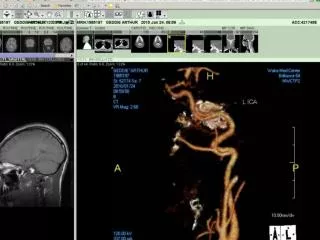
Cerebral Monitoring • Why is it important? • Once compromise is discovered (or predicted) carotid shunt can improve cerebral oxygen delivery • Carotid shunt can be placed in both external or internal carotid artery; however internal carotid is much more effective
Cerebral Monitoring • Why not shunt everyone? • Potential displacement of atheromatous debris, introduction of air embolism or thrombosis of shunt • Increases surgical time • Presence of shunt makes surgical field less than optimal
Cerebral Monitoring • Awake patient the gold standard • Assessment of grip strength of the contralateral hand • Responsive to verbal commands • Same anesthesiologist for assessment in comparison of before and after crossclamping
Cerebral Monitoring • Backpressure measurement • Gives an estimate of reasonable collateral circulation above the crossclamp • Carotid stump pressure to predict need for temporary shunt placement • Traditionally the cutoff has been 50 mmHg
Cerebral Monitoring • EEG current best measurement for GA patients • Gives ability to assess both focal and global changes • General anesthetic may change EEG patterns • Difficult to interpret, needing special expertise • BIS has been used to identify severe ischemia • Unable to differentiate global versus focal changes
Cerebral Monitoring • SSEP usefulness inconclusive • Retrospective review concluded could be useful • Prospective study of 50 patients concluded that although there is a 2% false negative rate, in general there is a limited value of SSEP in the detection of cerebral ischemia
Cerebral Monitoring • TCD ultrasonography noninvasive monitoring of the velocity of blood flow in the middle cerebral artery • Belardi suggests that U/S may not be effective in the prediction for shunt placement • Could be useful in the detection of cerebral emboli
Cerebral Monitoring • Carotid angiography may be a useful predictor of assessment of collateral circulation • Shunt more common when failure of collateral flow from contralateral hemisphere or when the contralateral internal collateral flow was occluded • Reported sensitivity 91% and specificity 35%