Download
1 / 30
310 likes | 355 Views
Cornea and sclera. cornea. Infective corneal lesions. Herpes simplex keratitis : Type 1 herpes simplex virus (HSV1) is a common imp. cause of ocular disease. Type 2 (HSV2) can cause genital disease may cause keratitis and infantile chorioretinitis .

E N D
Infective corneal lesions • Herpes simplex keratitis: • Type 1 herpes simplex virus (HSV1) is a common imp. cause of ocular disease. • Type 2 (HSV2) can cause genital disease may cause keratitis and infantile chorioretinitis . • Primary infection with (HSV1) aquired early in life by close contact such as kissing, it can be asymptomatic or with : • -fever • -vesicular lid lesion • -follicular conjuctivitis • -preauricularlymphadenopathy
The primary infection is followed by resolution and latency of the virus in the trigeminal ganglion , while the period of reactivation of the latent virus is “recurrent infection” in which the virus travels centrifugally to nerve terminals in the corneal epithelium to cause an epithelial keratitis. • The pathognomonic appearance is dendritic ulcer which is a linear , branching , epithelial ulcers
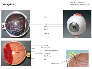
Corneal ulcer, or ulcerative keratitis, is an inflammatory or more seriously, infective condition of the cornea involving disruption of its epithelial layer with involvement of the corneal stroma.
May heal without scar or progress to a stromalkeratitis and inflammatory infiltration with perminant scar ,corneal grafting can be done . • Treatment: • Aciclovir • Ganciclovir • Vidarabine • triflurothymidine
Herpes zoster ophthalmicus • Varicella zoster. • Ophthalmic division of trigeminal nerve. • Pain and vesicles in the distribution of the ophthalmic nerve. • Ocular problems if nasociliary branch of the nerve is involved. • Signs: • Lid swelling • Keratitis • Iritis • Secondary glaucoma
Treatment: • Oral antiviral treatment for post infective neuralgia . • Antibacterials to prevent secondary infection . • Prognosis is improved with antivirals .
Bacterial keratitis • Staph.epidermidis ,staph.aureus , strep.pneumonia , coliform , pseudomonas , haemophilus. • Factors prevent infection of cornea and conjuctiva: • Blinking • Flow of tears • The corneal epithelium • Mucus trapping foreign bodies • Predisposing factors: • Keratoconjunctivitissicca • Contact lens wear • A break in the corneal epithelium
Signs and symptoms • Pain • Purulent discharge • Ciliary injection • Visual loss • Hypopyon • White corneal opacity
Treatment: • Gram staining and culture + topical antibiotics • Dual therapy to cover most bacteria (cefuroxime + gentamicin ) • Monotherapy ( ciproflpxacin ) • Tissue adhesives and corneal graft for perforated cornea
keratoconus • Painless disorder which is resulted from failure of cohesion between stromal collagen fibrils and lamellae of unknown cause, causing them to slip over one another and unravel. • —> resulting in progressive central corneal thinning –> leads to an ectatic conical cornea and myopia. • Mostly sporadic / inherited.
Presentation:young patient with myopia, irregular astigmatism , and in severe cases vision loss. • Diagnosis: 1- distorted red reflex during ophthalmoscopy . 2-record surface corneal topography.
Treatment: 1-rigid contact lenses arch over the irregularity of the cornea and restores the optics of the eye 2-replacement of the corneal stroma. 3-cross-linking of the anterior stromal collagen—> UVA radiation. 4-corneal graft.
Band shaped keratopathy • Subepithelial deposition of calcium phosphate in exposed part of the cornea —> co2 loss and consequent raised ph which favour its deposition. • Associated with: chronic uveitis, glaucoma, and systemic hypercalcaemia (hyperparathyrodism or renal failure).
Symptoms: 1-visual loss. 2-discomfort—>epithelial erosions. • Treatment: 1-symptomatic—> scraping off surgery with using off chelating agent such as sodium edetate. 2-excimer laser.
Lipid arcus • Asymptomatic. • Peripheral white ring lipid deposit. • Often Elderly people (arcus senilis) , if young >>hyperlipoproteinaemia. • No treatment required.
Episcleritis • Inflammation at the surface of the sclera. • Not associated with systemic diseases. • Symptoms: patches of redness and mild or no discomfort. • Treatment: 1-self limiting. 2-symptoms are tiresome—> topical anti inflammatory treatment. 3-severe —> NSAIDs
Scleritis ¶ More severe condition than episcleritis. ¶F>M , elderly ¶Collagen vascular diseases 50% , most commonly rheumatoid arthritis. It is a cause of deep ocular pain. Both inflammatory areas and ischaemic areas of the sclera may occur. Usually anteriorly. Characteristically the affected sclera is swollen. The following may complicate the condition: •Scleral thinning ( scleromalacia ), sometimes with perforation. • keratitis. •Uveitis. •cataract formation. •Glaucoma.
Treatment: 1- anti-inflammation treatment. 2-immunosuppressants. 3-steroids . 4-cytotoxic therapy. 5-sclera grafting—> prevention of perforation of globe.