Download
1 / 89
1k likes | 1.57k Views
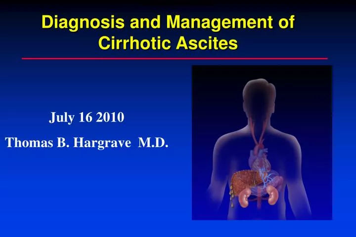
ASCITES AND HEPATORENAL SYNDROME. Diagnosis and Management of Cirrhotic Ascites. July 16 2010 Thomas B. Hargrave M.D. Complications of Cirrhosis. Ascites Hepatorenal Syndrome Spontaneous Bacterial Peritonitis (SBP) Hepatic Hydrothorax Varices Hepatic Encephalopathy (HE)

E N D
ASCITES AND HEPATORENAL SYNDROME Diagnosis and Management of Cirrhotic Ascites July 16 2010 Thomas B. Hargrave M.D.
Complications of Cirrhosis • Ascites • Hepatorenal Syndrome • Spontaneous Bacterial Peritonitis (SBP) • Hepatic Hydrothorax • Varices • Hepatic Encephalopathy (HE) • Hepatocellular Carcinoma (HCC)
Ascites • Ascites is the most common of the three major complications of cirrhosis • Approximately 50%-60% of compensated cirrhotics will develop ascites over 10 years of follow up • Development of ascites is an important landmark in the natural history of cirrhosis indicating a 50% 2-year mortality
Cirrhotic Ascites – Survival 100 80 60 Survival (%) 40 20 5 6 1 2 4 3 Onset Years
Wait List and Transplant Activity for Liver, 1998–2007 Waiting List at Year End Total Liver Transplants Deaths on Waiting List OPTN/SRTR Annual Report Tables 1.3, 1.6, 1.7
CIRRHOSIS IS THE MOST COMMON CAUSE OF ASCITES Cirrhosis is the Most Common Cause of Ascites Peritoneal malignancy Cirrhosis Heart failure 85% Peritoneal tuberculosis • Others • Pancreatic • Budd-Chiari syndrome • Nephrogenic ascites
Hepatic Sinusoid Unlike other capillaries, normal hepatic sinusoids lack a basement membrane. The sinusoidal endothelial cells themselves contain large fenestrae (200-400 nm in diameter), allowing passage of large molecules with molecular weight up to 250,000. These two features make the normal hepatic sinusoid very permeable with movement of fluid depending mostly on hydrostatic pressure Normal portal sinusoid pressure is 3-4 mmHg
Hepatic Sinusoid no basement membrane Hepatocytes The normal sinusoid is “leaky” Sinusoid Sinusoidal endothelial cells contain large fenestrae (200-400 nm in diameter), allowing passage of large molecules with molecular weight up to 250,000.
PORTAL VEIN OBSTRUCTION ALMOST NEVER LEADS TO ASCITES splanchnic capillary pressure Portal Vein Obstruction Almost Never Leads to Ascites Normal sinusoidal pressure Portal vein obstruction
HEPATIC VEIN OBSTRUCTION LEADS TO ASCITES FORMATION Splanchnic capillary pressure Hepatic Vein Obstruction Leads to Ascites Formation Hepatic vein outflow block Sinusoidal pressure
HVPG > 12 mmHg is Necessary for Ascites to Develop and is Associated with Low Sodium Excretion HVPG > 12 mmHg IS NECESSARY FOR ASCITES TO DEVELOP AND IS ASSOCIATED WITH LOW SODIUM EXCRETION 60 No ascites Ascites 50 40 Urinary sodium (mEq/L) 30 20 10 0 12 0 5 10 15 20 25 HVPG (mmHg) Morali et al., J Hepatol 1992; 16:249
NATURAL HISTORY OF ASCITES Natural History of Ascites Portal Hypertension No Ascites HVPG <12 mmHg Mild Vasodilation Uncomplicated Ascites HVPG >12 mmHg Moderate Vasodilation Refractory Ascites HVPG >12 mmHg Severe Vasodilation HVPG >12 mmHg Extreme Vasodilation Hepatorenal Syndrome
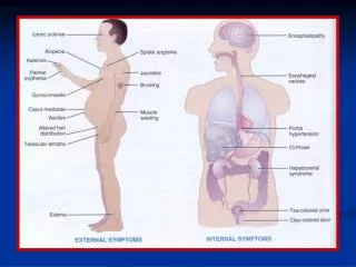
Ascites: Diagnosis • Physical examination is relatively insensitive in the diagnosis of moderate ascites • Sensitivity 50-94% Specificity 29-82% • Flank dullness is most specific sign • Approximately 1500 cc of fluid required for flank dullness • “Fluid wave” and “puddle sign” are not useful • Almost all patients with cirrhosis severe enough to develop ascites have stigmata of cirrhosis on physical examination
Ascites: Diagnosis • Ultrasound is the gold standard detecting as little as 100 cc. • Most cost -effective Ascites Liver
Ascites: Paracentesis • Abdominal paracentesis is the most rapid and cost-effective method of diagnosing the cause of ascites • Paracentesis should be performed in all patients with new-onset ascites and recently-admited inpatients with establishedascites • 20 % prevalence of SBP/infection at time of admission • Complications reported in only 1%, despite elevated INR in over 70% • Serious complications (hemoperitoneum or bowel entry) in <1/1000
DIAGNOSTIC PARACENTESIS Diagnostic Paracentesis Indications • New-onset ascites • Admission to hospital • Symptoms/signs of SBP • Renal dysfunction • Unexplained encephalopathy Contraindications • DIC
Ascites: Paracentesis • Coagulopathy should not preclude paracentesis unless there is clinically evident DIC • There is no data-supported cutoff or coagulation parameters beyond which paracentesis should be avoided. • Best location LLQ , 2 FB cephalad and 2Fb medial to ant. sup. Iliac crest.
ASCITES FLUID ANALYSIS Ascites Fluid Analysis Routine Optional • Albumin • Protein • PMN cell count • Cultures • Glucose • LDH • Amylase • Red blood cell count • TB smear and culture • Cytology • Triglycerides
Ascites: Analysis • For uncomplicated cirrhotic ascites, only three screening tests are initially indicated • Cell count • Albumin • Total protein • If SBP suspected, bedside bacterial cultures optimal • Serum-ascites albumin gradient (SAAG) is 97% accurate in diagnosis of portal hypertensive ascites • Cytology and mycobacteria cultures should only be ordered for a high index of suspecion.
Ascites Analysis The three main causes of ascites, cirrhosis, right-sided heart failure and peritoneal pathology (malignancy or tuberculosis), can be easily distinguished by combining the results of both the SAAG and ascites total protein content
The Serum-Ascites Albumin Gradient (SAAG) Correlates With Sinusoidal Pressure The serum-ascites albumin gradient (SAAG) is based on the fact that, per Starling forces, oncotic-hydrostatic balance is the major controlling force determining the protein concentration of fluid in the peritoneal cavity. The SAAG cutoff value that best distinguishes patients in whom ascites is secondary to liver disease and those with malignant neoplasm is a SAAG of 1.1 g/dL.
THE SERUM-ASCITES ALBUMIN GRADIENT (SAAG) CORRELATES WITH SINUSOIDAL PRESSURE 11 1.1 The Serum-Ascites Albumin Gradient (SAAG) Correlates With Sinusoidal Pressure 30 20 HVPG (mmHg) 10 r = 0.73 0 0 1.0 2.0 3.0 SAAG (g/dL) Hoefs J, J Lab Clin Med 1983; 102:260
SERUM-ASCITES ALBUMIN GRADIENT (SAAG) IS HIGH IN PORTAL HYPERTENSIVE CAUSES OF ASCITES Serum-Ascites Albumin Gradient is High in Portal Hypertensive Causes of Ascites 4.0 3.0 Serum – ascites albumin gradient (g/dL) 2.0 1.1 1.0 0 Cardiac ascites Peritoneal malignancy Cirrhotic ascites Runyon, Ann Intern Med 1992; 117:215
THE PERMEABILITY OF THE HEPATIC SINUSOID VARIES IN HEALTH AND DISEASE The Permeability of the Hepatic Sinusoid Varies in Health and Disease no basement membrane Hepatocytes Sinusoid The normal sinusoid is “leaky” fibrous tissue deposition “capillarization” of sinusoid In cirrhosis, the hepatic sinusoid is less leaky Sinusoid
Larger Serum Proteins Less Likely to Traverse the Sinusoid Fibrosis
ASCITES TOTAL PROTEIN LEVELS ARE ELEVATED IN CARDIAC ASCITES AND PERITONEAL MALIGNANCY 7.0 6.0 5.0 Ascitic fluid total protein (g/dL) 4.0 3.0 2.5 2.0 1.0 0 Cardiac ascites Peritoneal malignancy Cirrhotic ascites Ascites Total Protein is Elevated in Cardiac Ascites and Peritoneal Malignancy Runyon, Ann Intern Med 1992; 117:215
SERUM-ASCITES ALBUMIN GRADIENT (SAAG) AND ASCITES PROTEIN LEVELS IN THE MOST COMMON CAUSES OF ASCITES (75) Serum-Ascites Albumin Gradient and Ascites Protein Levels in the Most Common Causes of Ascites 4.0 3.0 Serum – ascites albumin gradient (g/dL) 2.0 1.1 1.0 0 Cardiac ascites Peritoneal malignancy Cirrhotic ascites 7.0 5.0 Ascitic fluid total protein (g/dL) 3.0 2.5 2.0 0 Runyon, Ann Intern Med 1992; 117:215
ASCITES CAN BE CHARACTERIZED BY SERUM-ASCITES ALBUMIN GRADIENT (SAAG) AND ASCITES PROTEIN Ascites Can Be Characterized by Serum-Ascites Albumin Gradient (SAAG) and Ascites Protein Source of ascites SAAG > 1.1 Hepatic sinusoids SAAG < 1.1 Peritoneum Ascitesprotein < 2.5 “Capillarized” sinusoid Ascites protein > 2.5 Normal “leaky” sinusoid Ascites protein > 2.5 Peritoneal lymph • Sinusoidal hypertension • Cirrhosis • Late Budd-Chiari • Post-sinusoidal hypertension • - Cardiac ascites • Early Budd-Chiari • Veno-occlusive disease Peritoneal pathology - Malignancy - Tuberculosis
Ascites: Additional Tests • Amylase • Pancreatic ascites • Glucose: low when being consumed by bacteria or WBC • Carcinomatosis • Gut perforation • Gram stain: limited sensitivity 7-10% • Required 10,000 bacteria/ml (medial bacterial concentration in SBP= 1/ml
Ascites: Additional Tests • Cytology • 100% sensitive for carcinomatosis • Only 2/3 of malignant ascites have carcinomatosis • Hepatocellular carcinoma • Massive liver metastasis • Chylous ascites due to lymphoma • Useless tests: • pH • Lactate • CEA
Ascites: Treatment • Successful treatment depends upon accurate diagnosis of the cause • Diuretics/sodium restriction ineffective in malignant, nephrogenic, and pancreatic ascites • If possible, treat underlying cause of the decompensated cirrhosis • ETOH: abstinence • Autoimmune Hepatitis: steroids, AZA • Hepatitis B: antivirals
NEUROHUMORAL SYSTEMS ARE ACTIVATED IN CIRRHOTIC PATIENTS WITH ASCITES Neurohumoral Systems are Activated in Cirrhotic Patients with Ascites Plasma renin activity (ng/ml-h) Plasma aldosterone activity (ng/dl) 70 800 700 60 600 50 500 40 400 30 300 250 20 200 150 10 100 50 0 0 Controls Cirrhosis no ascites Cirrhotic patients with ascites Controls Cirrhosis no ascites Cirrhotic patients with ascites
Ascites: Treatment • Goals: • Minimize ascitic volume and peripheral edema • Avoid intravascular volume depletion • Benefits • Patient comfort • Reduced risk of hernia formation • Possible reduction in SBP due to increased concentration of ascitic fluid opsonins • Improved nutrition
Treatment: Create Negative Sodium Balance • The typical patient with ascites excretes < 20 meq sodium/day • The typical North American diet contains 200-300 meq sodium/day • No-added salt diet = 100-150 meq sodium • A diet containing 88 meq sodium/day is the best compromise between salt restriction and acceptable palatability
Treatment: Create Negative Sodium Balance • Dietary sodium restriction is essential to effective management of ascites • 140 meq sodium = 1 liter of retained fluid • Fluid loss follows sodium loss, so fluid restriction is rarely necessary • Hyponatremia is common with ascites and does not require treatment if the Na > 130 meq/l • The vast majority of patients require diuretics
Spironolactone Furosemide
Treatment: Diuretics • The most successful regiment is a combination of a single morning oral dose of • spironolactone 100mg • furosemide 40 mg • Maximizes compliance and minimizes nocturia • The absorption of spironolactone is improved if given with food
Treatment: Diuretics • Both proximal and distal acting diuretics needed to achieve diuresis • Half life 4-6 hours • Furosemide is filtered in the glomerulus and blocks sodium reabsorption on the luminal side • A concentration-dependent threshold effect is observed
Rational Use of Furosemide Furosemide half-life 4-6 hours Luminal Concentration Furosemide Diuretic Threshold Furosemide 40 mg 8 PM 8 AM
Rational Use of Furosemide Furosemide half-life 4-6 hours Luminal Concentration Furosemide Diuretic Threshold Furosemide 40 mg Furosemide 40 mg 8 PM 8 AM
Rational Use of Furosemide Luminal Concentration Furosemide Diuretic Threshold Furosemide 80 mg 8 PM 8 AM
Treatment: Diuretics • The diuretic effect of spironolactone is seen within 48 hours and peaks at 2 weeks • Dose should be adjusted no more often than once every 5-7 days • Doses can be doubled in the absence of diuresis to a maximum of spironolactone 400 and furosemide 160 mg/day • The spironolactone/furosemide ratio of 100/40 should be maintained to maintain normokalemia
Untreated Urine Lytes NA < 10 mEq/K> 40 mEq Urine Electrolytes and Treatment Decisions Inadequate Diuresis 2 gram (88 mEq) sodium diet Furosemide 40 mg + Spironolactone 100 mg NA = 20 mEq K= 20 mEq NA = 40 mEq K= 60 mEq NA = 100 mEq K= 20 mEq Discuss Dietary Compliance Increase Spironolactone Increase Furosemide
Treatment: Diuretics • Two major concerns • Avoid overly rapid diuresis: pre-renal azotemia • Progressive electrolyte imbalance • Rate of safe diuresis is a function of presence or absence of peripheral edema • Peripheral edema = rate unlimited • The maximum rate of mobilization of ascites via peritoneal capillaries is 500-700 cc/day • Weight loss should not exceed 0.5 kg/day