Download
1 / 22
220 likes | 300 Views
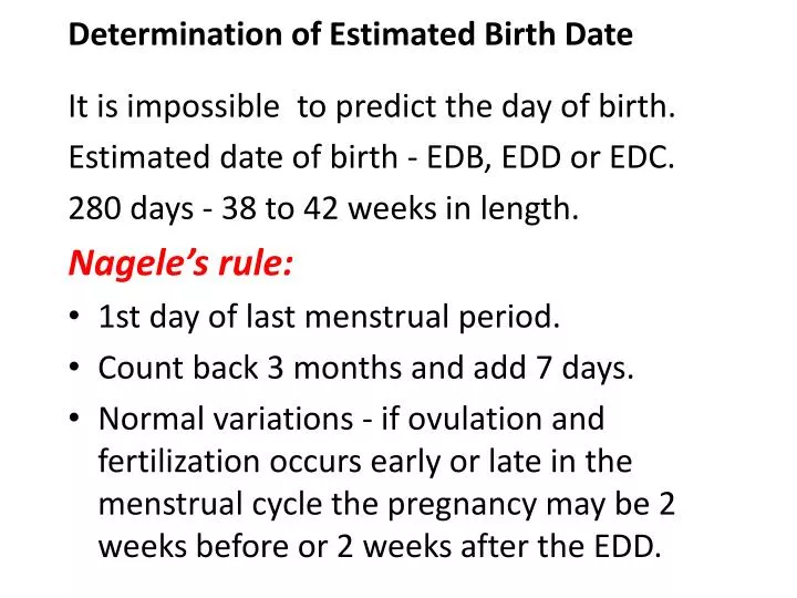
Determination of Estimated Birth Date. It is impossible to predict the day of birth. Estimated date of birth - EDB, EDD or EDC. 280 days - 38 to 42 weeks in length. Nagele’s rule: 1st day of last menstrual period. Count back 3 months and add 7 days.

E N D
Determination of Estimated Birth Date It is impossible to predict the day of birth. Estimated date of birth - EDB, EDD or EDC. 280 days - 38 to 42 weeks in length. Nagele’s rule: • 1st day of last menstrual period. • Count back 3 months and add 7 days. • Normal variations - if ovulation and fertilization occurs early or late in the menstrual cycle the pregnancy may be 2 weeks before or 2 weeks after the EDD.
Fetal Growth and Development • Nurses responsibility • signed consent form with information on the procedure and possible risks • scheduling the procedure • explaining the procedure to the woman and support person • preparing the woman physically and psychologically • providing support during the procedure • assessing both fetal and maternal responses to procedures • provide follow up care • manage equipment and specimens
Fetal Growth and Development Estimated Fetal Growth: • McDonald’s rule - method of determining, during mid pregnancy, that the fetus is growing in utero by measuring fundal (uterine) height. • Distance from fundus to symphysis in centimeters is equal to the week of gestation between week 20 to 31. • Measure from the notch of the symphysis pubis to over the top of the fundus with woman lies supine. • This becomes inaccurate in 3rd trimester because the fetus is growing in wt. than height. • Milestones: • 12 weeks- over symphysis pubis • 20 weeks at umbilicus • 36 weeks xiphoid process
Fetal Growth and Development Assessing Fetal Well-Being: • Fetal movement - quickening week 18 to 20 • peaks at week 28 to 38 • healthy fetus moves 10 times a day. • Ask mother to lie in left recumbent position after a meal and record number of fetal movements in next hour. • 2 times in 10 minutes • fewer than 5 in 1 hour - notify the doctor.
Assessing Fetal Well-Being Fetal Heart Rate: • 120 to 160 beats per minute throughout pregnancy. • Week 10 to 11 heart sounds can be heard and counted with a doppler. Rhythm Strip Testing: • baseline fetal heart rate per minute and long and short term variability. • Place woman in semi-fowlers (prevents supine hypotension syndrome).
Assessing Fetal Well-Being • Monitors are attached abdominally and recorded for 20 minutes (mother in fixed position) • Short term variability- beat to beat variability- denotes small changes in rate from second to second if parasympathetic nervous system is receiving adequate O2 and nutrients. • Long term variability - denotes the differences in heart rate that occur over 20 minutes (fetus moves 2x/10 min) increases with movement.
Assessing Fetal Well-Being • This reflects fetal sympathetic nervous system. Nonstress Testing: • measures response of fetal heart rate to movement. • Monitors are attached to abdomen • mother pushes a button attached to the monitor when she feels the fetus move. • FHR should increase 15 beats/ min and remain elevated for 15 seconds.
Assessing Fetal Well-Being • Decreases when fetus quiets. If no increase is noticeable with movement poor O2 perfusion of fetus id suggested. • Test lasts 10 to 20 minutes. • If no fetal movements in 20 minutes fetus may be sleeping. • Orange juice or carbohydrate may increase blood glucose level which stimulate the fetus. Also loud noise may stimulate fetus.
Assessing Fetal Well-Being Vibroacustic Stimulation: • Acustic stimulation (artificial larynx) applied to abdomen to produce a sharp sound, startling and waking the fetus. • 80 dB frequency of 80 Hz. Contraction Stress Testing: • FHR is analyzed in conjunction with contractions. • Mother stimulates the nipple which releases oxytocin which initiates uterine contractions
Assessing Fetal Well-Being • External uterine contraction and FHR monitors are applied • 3 contractions with duration of 40 seconds or more present in a 10 minute period. • Normal (negative) when no FHR decelerations are present with contractions. • Abnormal (positive) 50% or more contractions cause a late deceleration (dip in FHR) toward the end of a contraction and continues after the contraction. • Woman waits 30 min after the test.
Assessing Fetal Well-Being Ultrasound: • response of sound waves against objects. • Diagnose pregnancy at 6 weeks gestation • confirm presence, size, and location of placenta and amniotic fluid. • Establishes fetal growth, gross defects • Establish presentation and position (sex) • Predict maturity by biparietal diameter • Mother has to have a full bladder ( drink a full glass of water q 15 min. in 1 1/2 hours
Assessing Fetal Well-Being • Place a towel under the right buttock to tip uterus away from the vena cava. • Gel applied to abdomen (room temperature) • Transducer is applied intravaginal or abdominal • Picture of sonogram Biparietal Diameter: • measures side to side measurement of fetal head (8.5cm or more infant weighs > 2500g 5.5 lb) 40 week gestation.
Assessing Fetal Well-Being • Also measures head circumference and femoral length. Doppler Umbilical Velocimetry: • Measures velosity at which RBC and vessels are traveling. Placental Grading: • amount of calcium deposits in base of placenta. Amniotic Fluid Volume Assessment: • average index is 15 cm between 28-40 wks.
Assessing Fetal Well-Being ECG at week 11 of pregnancy (inaccurate before week 20 because fetal electrical conduction is week). MRI used to diagnos ectopic pregnancy or trophoblastic disease. Maternal Serum Alpha-Fetoprotein is a substance produced by the fetal liver that is present in amniotic fluid and maternal serum. Begins to rise at week 11. • Detects Down Syndrome, open spinal or abdominal defects.
Assessing Fetal Well-Being Triple Screening - analysis of 3 indicators: • Maternal serum Alpha-fetoprotein • unconjugated estriol • hCG • used for Downs syndrome Chorionic Villi Sampling (CVS) • biopsy and analysis for chromosomal analysis done at week 10 to 12.
Assessing Fetal Well-Being Amniocentesis: • aspiration of amniotic fluid from the uterus for examination. • Week 12 to 13 • 1 mL of fluid is needed • 3 to 4 in 20 to 22 gauge spinal needle • woman rest for 30 minutes after the procedure • constant monitoring for FR and contractions • if Rh-neg. blood give RhoGAM
Assessing Fetal Well-Being Amniocentesis: • color of water or slight yellow tinge • strong yellow- bilirubin • green- meconium • lecithin/ sphingomyelin ratio • protein components of lung enzyme surfactant that alveoli form week 22-24 • phosphatidyl glycerol and desaturated phosphtidylcoline • found in surfactant
Assessing Fetal Well-Being • Bilirubin Determination • must be blood free to analyze bilirubin • Chromosome Analysis • uses fetal skin cells for karyotyping • Fetal Fibronectin • glycoprotein that helps placenta attach to the uterine decidua ( preterm labor). • Inborn Errors of Metabolism • inherited diseases from inborn errors
Assessing Fetal Well-Being • Alpha-Fetoprotein Percutaneous Umbilical Blood Sampling • aspiration of blood from the umbilical vein for analysis Amnioscopy • visual inspection of amniotic fluid through the cervix and membranes with a small fetoscope (detects meconium). Fetoscopy • visualizing the fetus with fetoscope
Assessing Fetal Well-Being • confirms intactness of spinal column • biopsy of fetal tissue and blood sample • surgery • photos • done week16 to17 at the earliest • risks-premature labor, infection .
Assessing Fetal Well-Being Biophysical Profile (fetal Apgar) • combines 4 to 6 parameters into one assessment • fetal movement and breathing, fetal tone, amniotic fluid volume, placental grading fetal heart reactivity. • more accurate in predicting well being than any single assessment. • a score of 4 to 6 denotes a fetus in jeopardy.