Download
1 / 30
320 likes | 486 Views
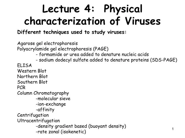
Lecture 4: Physical characterization of Viruses. Different techniques used to study viruses: Agarose gel electrophoresis Polyacrylamide gel electrophoresis (PAGE) - formamide or urea added to denature nucleic acids - sodium dodecyl sulfate added to denature proteins (SDS-PAGE) ELISA

E N D
Lecture 4: Physical characterization of Viruses Different techniques used to study viruses: Agarose gel electrophoresis Polyacrylamide gel electrophoresis (PAGE) - formamide or urea added to denature nucleic acids - sodium dodecyl sulfate added to denature proteins (SDS-PAGE) ELISA Western Blot Northern Blot Southern Blot PCR Column Chromatography -molecular sieve -ion-exchange -affinity Centrifugation Ultracentrifugation -density gradient based (buoyant density) -rate zonal (isokenetic)
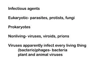
Isolation and Detection, and Measurement of Viruses • Isolation. Must first be isolated from a source, e.g. • whole organism or a part thereof • excreted or secreted material, • blood, • tissue. • Samples are then typically processed. • Detection can be based on numerous methodologies. • Clinical: the manifestation of some abnormality in host organisms or host cells. • Epidemiological: Clinical but on the scale of populations. • Diagnostic: Involves a test to physically detect the presence of virus.
Measurement of Viruses Measurement: Physical and functional methods to enumerate viruses. • Physical Methods: Electron microscopy, optical density, Hemagglutination assay, various immunoassays (e.g. ELISA, RIA), quantitative PCR. • Functional Methods...the Infectious Unit: the number of viral particles it takes in order to establish an infection
Common techniques applied in Virology for studying viruses and viral components Technique • Separation of proteins and nucleic acids by size • Agarose gel electrophoresis • Polyacrylamide gel electrophoresis (PAGE) • PAGE + Urea or Formamide • PAGE + SDS (sodium dodecyl sulfate) • Detection of nucleic acids by autoradiography • Northern Blot (RNA) • Southern Blot (DNA) • PCR • Immunodetection • Western Blot • ELISA (Enzyme-linked immunosorbant assay)
Common techniques applied in Virology for studying viruses and viral components Technique • Column Chromatography (gel filtration or molecular sieve) • ion-exchange • affinity • Centrifugation • in aqueous buffer • rate zonal or isokenetic • buoyant density or isopycnic • Electron microscopy
Centrifugation • Ultracentrifuge- A centrifuge capable of generating large centrifugal fields by rotating samples at 20,000-100,000 rpm. Centrifugal forces of greater than 100,000 X gravity can be generated. • Sedimentation coefficient • Rate at which a macromolecule sediments under a defined gravitational force. • Influenced by • molecular weight • shape of a macromolecule • The basic unit is the Svedberg (S):10-13 sec. • Can be used to estimate molecular weights in conjunction with other values. • Buoyant density-Density at which a virus or other macromolecule neither sinks nor floats when suspended in a density gradient (e.g., CsCl2 or sucrose).
The Svedberg equation S= Sedimentation coefficient v = velocity w = angular velocity (in radians/sec. 1 revolution = 2p radians) r – radius, i.e. distance from center of rotation m = mass (grams) = partical specific volume of particle (in nm) r = density of solvent (g/cm3) f = frictional coefficient between particle and solvent. For a globular protein, f ≈ 1 (fp = frictional coeffieient of the particle; fm = frict. coeff. of solvent). = volume of the particle
Sedimentation media • Aqueous Buffer (Water based)- • Used to separate molecules with widely different S values (ex. Nuclei from ribosomes) • Sucrose or glycerol gradients or cushions (isokenetic or rate-zonal)- • Fixed concentration or linear gradients. • Increase the density and viscosity • Decreasing sedimentation rates • preventing the sedimentation molecules with densities less than the medium. • Controlling the time and speed of centrifugation a significant purification can be obtained. • Since most macromolecules have greater densities than these mediums separation is based on S values. • Can be used to separate molecules with relatively close S values.
Sedimentation media • CsCl gradient centrifugation (isopycnic or buoyant density)- • Linear gradient of these compounds in buffer is prepared in the centrifuge tube. • As the concentration of the compound is increased the density of the medium increases in the tube. • Density is low at the top and high at the bottom. • Macromolecule centrifuged through will form a band at a position equal to buoyant density. • Useful for separating molecules of different densities even when the densities are very close. • Drawback: CsCl can permanently inactivate some viruses.
Other techniques for separation Electrophoresis, column chromatography. • Not normally used to separate virions but are used to separate nucleic acids or proteins. • Can be used to separate and purify virions in some cases. • Both methods separate macromolecule on the basis of charge and/or size characteristics. • Although virions have a variety of charged macromolecule only those charged groups exposed to the surface contribute to electrophoretic mobility or ion-exchange characteristics. • Chromatography can be ion exchange in which charged groups are ligated to the chromatographic medium. • The charged virus can then be bound to the medium and eluted by increasing the concentration of a competing ion. • Molecular sieve chromatography allows for very large pores to be formed which virus particles can enter
Electrophoresis Size exclusion chromatography
Indirect detection of viral nucleic acids • Blots: after gel separation, transfer to solid phase and probe with labeled nucleic acids • Southern Blot: detects DNA • Northern Blot: detects RNA Southern blot (Fig 2.13)
Direct Detection of viral nucleic acids • PCR based assays: Directly amplifies nucleic acids from sample • Microarray technology: examines the effects of viral infection on gene expression
Saiki, et al. 1988. Science 239: 487-491. Revolutionized the molecular biosciences as we know them.
2n - 2(n-1) -2 duplex DNA molecules of the expected length after n cycles of amplification (n = 1, 2, ...) PCR- the method
Assessing the purity of virions Spectrophotometric analysis UV absorption at 260 and 280 nm. This ratio (260/280) is a characteristic of a pure virus and is dependent on the amount of nucleic acid and protein in the virus. The number can be used to estimate the amount in the preparation. Nucleic acid absorbs light about twice as well at 260 vs. 280 and vice versa for protein.
Assessing the purity of virions Serological methods • Antibodies to viral proteins are used to: • Characterize • Detect • Quantify virions.
Antibodies can be made in several ways. • Whole virus or recombinant proteins injected into animals • Polyclonal, monoclonal • Recombinant single chain antibodies made in E. coli.
Use of Antibodies: direct and indirect detection of viral antigen. (Fig. 2.8)
Methods for using antibodies • ELISA (Enzyme-linked immunosorbent assay), • RIA (radioimmune assay) • RIPA (radioimmune precipitation assay) • Colorometric bead-based diagnostic assays • Western blotting • Direct precipitation of virus with antibody • Neutralization of viral infectivity • Complement fixation by the virus-antibody complex
ELISA: Enzyme Linked Immuno Sorbant Assay To detect viral proteins in a sample Fig. 2.11 To detect antibodies to a viral protein
Electron microscopy • Allows the visualization of single virus particles. • Resolution in the nm range (10-9 meters) is possible. • Based on the principle of electron scattering. • A beam of electrons is focused on the sample. Electrons within the specimen will scatter the electron beam. • The scattering effect is enhanced by the presence of heavy, electron rich metal ions within the sample. • SEM (Scanning EM) versus TEM (transmission EM) • Negative staining: stains background but not the virus particles) • Shadowing techniques. Creates a region where relatively little metal deposits just behind the viral particle (resulting in a shadow). • Cryo-EM: freeze particles in vitreous ice, do SEM, collect images, and reconstruct in 3-D
T4 phage Negative staining TEM of HIV
Cryo-EM: freeze particles in vitreous ice, do SEM, collect images, and reconstruct in 3-D
X-ray crystallography- • Analysis of crystallized virus. • Virus crystals are symmetrical structures composed of many isometric viruses. • Atoms of the crystal diffract X-rays in a structure dependent manner. • Resolution at the Å level (10-10 meters, in the bond length range) is possible. • This approach has been used to analyze the structure of the viruses at the molecular level.