Download
1 / 12
130 likes | 393 Views
Cytoskeleton. Gives the cell mechanical strength, controls its shape, drives and guides its movement Four classes Muscle thick filament Microfilaments Intermediate filaments Microtubules. Changeable cytoskeleton. The cytoskeleton changes in response to its environment

E N D
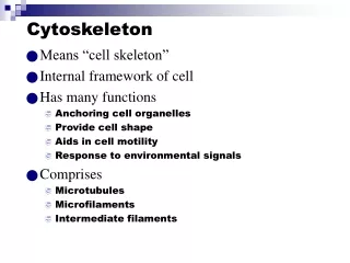
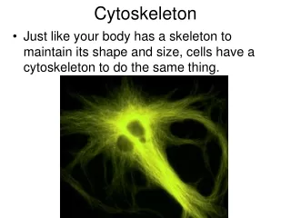
Cytoskeleton • Gives the cell mechanical strength, controls its shape, drives and guides its movement • Four classes • Muscle thick filament • Microfilaments • Intermediate filaments • Microtubules
Changeable cytoskeleton • The cytoskeleton changes in response to its environment • Are there chemicals the cell likes to stick to? • Is the surface flat or bumpy? • Is the surface stiff or soft? • Are the cells near other cells?
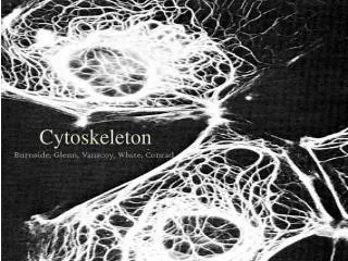
Where is actin in a cell? Contractile ring In dividing cells Microvilli cores And terminal web Leading edge of Motile cell Stress fibers And cell cortex
Lamellipodia, filipodia and blebs Pinocytotic cup lamellipodia • Membrane protrusions under the control of actin • Can view these as cells move across a surface Bridge still connecting recently divided cells filipodia bleb
Solutions and Equipment • PBS • Used to wash the cells • 4% Formaldehyde • Preserves the cells • Glycine • Will prevent other chemicals in the cells from glowing • 0.4% Triton X-100 • Pokes holes in the membrane so the stain can go in
Rhodamine Phalloidin • Phalloidin • From the poisonous mushroom Amanita phalloides (Death Cap) • Binds specifically to actin • Rhodamine • Fluorescent marker stuck to phalloidin • When we shine green light on the sample (l = 520-560 nm) • The sample will glow red (l = 650-720nm)
Mounting Media • Helps the coverslip stick to the slide
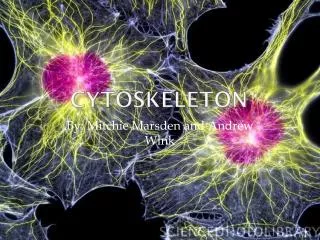
Fluorescence Microscopy • Green light photons “promote” rhodamine electron to a higher energy level. When they return to original level, they emit a lower energy photon. • What you can expect to see • Black background, with red stress fibers visible
Photobleaching • Keep sample protected from light after adding rhodamine phalloidin • If any fluorescent sample is left exposed too long to light, the fluorescence will fade • Results will not be as good
Multi colored fluorescence images require changing excitation sources and layering the images • Blue is the nucleus • Red is actin • Green is golgi • Pink is mitochondria