Download
1 / 26

E N D
Lymphatic System Human Anatomy Chapter 20
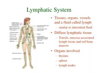
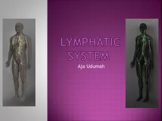
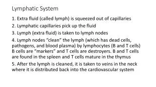
I. The lymphatic system- This system involves a set of vessels that transports tissue fluid back into the blood stream and a set of organs that are involved in the immune system. The lymph vessels are as branched and abundant as arteries and veins. They collect lymph- tissue fluid arising from fluid that has leaked from the capillaries. Proteins also leak from capillaries and are returned to the blood system by the lymph vessels. The lymphatic system also collect lipids from the digestive tract and is involved in exposing pathogens to the immune system.
A. lymph capillaries- one cell layer thick made up of endothelial cells that are permeable to tissue fluid, proteins, bacteria, and viruses. Lymph capillaries are not located in bone, bone marrow, teeth, and the central nervous system. Lacteals are lymph capillaries in the small intestine that are permeable to lipids.
B. lymphatic collection vessels- These vessels connect to the capillaries and carry the fluid away. Their tunic structures are like those of blood vessels but overall they are thin and difficult to identify. They contain valves as veins do but unlike veins they contain bulges making the vessel resemble a beaded string.
C. lymph nodes- organs along the lymphatic collecting vessels that accumulate the pathogens carried by lymph. This is an area highly concentrated with lymphocytes ready to activate an immune response. D. lymph trunks- a conversion of lymphatic collection vessels that results in a vessel of a larger diameter. They drain large areas of the body. Five major locations areL lumbar trunks, intestinal trunks, bronchomediastinal trunks, subclavian trunks, and jugular trunks. E. lymph ducts- The larges vessel into which the lymph trunks empty. The thoracic duct is found in everyone but only some people have the right lymphatic duck that empties into the neck veins. The thoracic duct empties into the left internal jugular and left subclavian veins.
A look inside the human lymphnode: the removal of most lymphocytes reveals the reticular tissue network.
II. The Immune system- This is a very complex system with multiple layers and forms of defense. In this section we focus on the organs closely associated with cellular response of the immune system. The immune system identifies and attacks specific pathogens. A. Lymphocytes and other cells of the immune system- Inflammation may be the first response to infectious organisms entering the body. Cells involved in an immune response are macrophages and lymphocytes. These include T and B cells. WBCs can move in and out of the blood vessels and also travel through lymph tissue in order to fight disease. 1. activation of lymphocytes- lymphoid stem cell in bone marrow give rise to lymphocytes. As they mature they are “taught” what tissue or cells are “self”, anything deviating from this, is a pathogen and will be “attacked”. a. Lymphocytes mature in the thymus (T-cell) or in the bone marrow (B-cell). b. Once they are mature they travel through the body until they encounter an antigen. c. Cells such as macrophages and dendritic cells engulf pathogens and present antigens to lymphocytes. d. A T-helper cell is activated once presented with antigen and it sends signals to activate other T-cells and the B-cells. e. Once activated the cells produce effector cells that fight the pathogen and memory cells. Memory cells guard against subsequent infections.
B. Lymphoid tissue- this is located throughout the body and is often infected because it is designed to collect pathogens for the purpose to destroying them. At the site of infection multiple lymphocytes collect to fight infection and memory cells reside. This tissue is located in areas frequently exposed to pathogens such as: mucous membranes of digestive, respiratory, urinary, and reproductive tracts. Also in the lymphoid organs which easily collect pathogens.
C. Lymphoid organs 1. lymph nodes- organs that filter lymph fluid before it has an opportunity to enter back into the blood vessels. It also collects antigens that are funneled to lymph tissue so they can engulf by macrophages and presented to lymphocytes. They are a cortex and medulla. .
2. spleen- largest lymphoid organ, it is almost the size of the heart, it is in the superior left abdominal area. It is designed to remove blood born pathogens and aged/damaged RBCs. It also stores platelets. It has areas identified at white (lymphoid tissue) and red pulp (vacular tissue).
3. thymus- lies posterior to the sternum in superior thorax, site to T-cell development. It also releases hormones. It is gradually replaced by fibrous tissue as one ages. It also has a cortex and medulla.
4. tonsils- simplest lymphoid organs, humans have four groups: palatine, lingual, pharyngeal, and tubal. They gather pathogens entering through the nose and mouth
5. aggregated lymphoid follicles and the appendix- densely packet units of lymphoid tissue forming lymphoid nodules, they are especially around the appendix and distal part of the small intestine, and in the cecum of the large intestine.
D. HIV virus and AIDS 1. HIV is transmitted through bodily secretions such as blood,semen and viginal secretions. If the fluid is exposed to air it dries, the virus dies and cannot be infectious. Any microscopic tear in the body can allow the virus entry if one is exposed to an infected body fluid. Entry can also occur via a needle or blood transfusion (rare). 2. The virus targets T-Helper cells and destroys them. It has the ability to incorporate itself into the cell’s genome and to mutate rapidly such that the immune system has a hard time keeping up with it. It also affects microglia cells (brain tissue) and immune system cells (dendritic and macrophages).
3. The infection stages are a. Acute stage (2 weeks): first infected presents flu-like symptoms, fever, rash, fatigue, headache, muscle/joint pain, diarrhea, swollen lymphnodes b. Asymptomic period (up to 10 years): there are no obvious symptoms but the immune system is silently fighting the HIV virus c. AIDS (time varies but it results in death): The persons’s immune system declines and the body is invaded by opportunistic organisms. AIDS patients have infections not seen in people with healthy immune systems. 4. The virus has the ability to quickly mutate and many of the treatments developed, like the cocktails, are not efficient. The number of people newly infected puts AIDS at the fourth leading cause of death world wide. It most severe spread is in Asia, Eastern Europe, and Africa. In the US 25% of people infected do not know they are. There is no absolute prevention of spreading infection other than abstinence. (Hint- don’t have sex with a partner until they take an AIDs test- remember the test can give a false positive for six-months after newly infected)