Download
1 / 23
230 likes | 353 Views
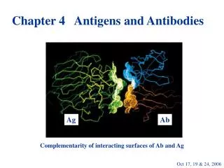
Lecture 2 ANTIGENS AND THEIR PROCESSING 2013/2014. ANTIGENS. The structure of antigens Antigen processing and presentation Peptide-MHC molecule: structure and assembly Antigen recognition by B and T cells Superantigens Stress proteins (heat shock proteins).

E N D
ANTIGENS The structure of antigens Antigen processing and presentation Peptide-MHC molecule: structure and assembly Antigen recognition by B and T cells Superantigens Stress proteins (heat shock proteins)
TYPES OF ANTIGENS INVOLVED IN PATHOGENESIS OF DISEASES • Microbial antigens (bacterial, viral, fungal, parasitic ones) • Blood group antigens • Transplantation alloantigens (MHC and minor ones) • Allergens • Autoantigens (organ and/or tissue specific) • Tumor antigens (tumor specific and tum. associated) • Superantigens: eg.SEB-staphylococcal enterotoxin B • Heat shock proteins
ANTIGENS FOR B CELLS • Antigens contain epitopes that can bind to the antigen-binding sites of antibodies • Antigens (haptens) may have almost any chemical nature • Antigens on native proteins are usually discontinous segments of aminoacids at cell surface • Antigens as immunogens must contain carrier epitopes for activating helper T cells
Antigen presenting cells (APC) • Professional APC: dendritic cells – the only ones which can stimulate naive T cells, • Activated macrophages, • B lymphocytes – ingest protein antigens and display them to helper T cells • All nucleated cells can function as APC, if possess foreign, usually microbial antigens in the cytoplasm following infection
ANTIGEN PROCESSING AND PRESENTATION FOR CLASS I MHC • Peptides presented on most cell types are synthetized endogenously, • Peptides presented by professional APC can be acquired through endocytosis, • Peptides are processed by proteasomes and enter endoplasmic reticulum through TAP transporter, • Peptides are bound at both termini within the binding cleft, • Antigens presented by class I MHC molecules are recognized by CD8 T cells
ANTIGEN PROCESSING AND PRESENTATION FOR CLASS II MHC • Only professional APC (dendritic cells, macrophages and B cells) express class II MHC constitutively • Peptides presented by professional APC are acquired mostly by receptor-mediated endocytosis, or pinocytosis • Peptides presented by class II molecules often have terminal extensions • Antigen presented by class II MHC molecules are recognized by CD4 T cells
Cross-presentation (cross-priming) • If infected cells and their viral antigens are ingested by APCs and broken down in APC cytoplasm, • If APC itself is infected • APC then acts as any nucleated cell, using proteasome pathway and presents peptides via MHC class I to CD8+ CTL • The same APC may display peptides via MHC class II to CD4+ T helper cells
Why and what for is the cross-presentation? • About 25 % of class I molecules present antigens of exogenous origin, • Up to 20% of MHC class II molecules present peptides derived from either cytoplasmic or nuclear antigens, • Naive cytotoxic T cells require dendritic cells for their activation,but most viruses are not tropic for DC and thus are not usually present in the cytosol of APC. • This is solved by sneaking out of the vacuole containing ingested external antigens to the cytosol , • Similarly, proteasome-derived peptides are taken up by so-called autophagosomes, by the mechanism of autophagy. The fusion with MHC class containing MIIC, where proteolytic cleavage of any intact proteins may also take place.
MAJOR HISTOCOMPATIBILITY (MHC) ANTIGENS • Histocompatibility antigens are cell surface expressed on all cells (class I) and on APC, B cells, monocytes/macrophages (class II) • Their physiologic function is to display peptides derived from protein antigens to antigen-specific T lymphocytes
MAJOR HISTOCOMPATIBILITY (MHC) ANTIGENS (2) • They are targets for graft rejection • They are inherited from both parents as MHC haplotypes and are co-dominantly expressed • They exist in multiple alleles (variants) distinct in particular individuals
Features of peptide binding to MHC molecules • Each MHC molecule displays one peptide – self if not infected • Many different peptides can bind to the same MHC molecules • Peptides have very slow off-rate • Stable expression requires bound peptide • MHC molecules bind only peptides
Nonclassical MHC molecules • They include HLA-E, HLA-F, HLA-G, MICA, MICB, HFE • They may be precursors to classical MHC ones • They are expressed on various cells but most often in gastro-intestical tract • Some have well- defined function: HLA-G expressed on placental-maternal trophoblast interface, protect conceptus from destruction by NK cells. The latter possess ILT2 inhibitory receptor that recognize HLA-G.
The family of CD1 non-MHC molecules • Encoded by a set of genes on chromosome 1 • CD1 is also involved in the presentation of Ag to T cells,l but it Ag-binding groove contains mainly hydrophobic aminoacids and its entrance is narrow • CD1 molecules present lipids or glycolipids but not proteins • There are four CD1 molecules on human cells – CD1a, b, c (on cortical thymocytes, dendritic cells) CD1d – on G-I tract, hepatocytes, lymphoid and myeloid cells.
ABO BLOOD GROUP ANTIGENS • ABO locus – encodes a glycosyl transferase and has three alleles: A, B (alloenzymes), O – functionally silent • O-individuals make anti-A and anti-B antibodies due to exposure to common bacteria • O-individuals are universal donors because their anti-A,B Abs bind to so many different cells in the recipient that they are effectively diluted • A and B-individuals may not donate blood to an O recip. because their erythrocytes will be lysed by anti-A and anti- B Ab
PROPERTIES OF SUPERANTIGENS • Presented and recognized as anunprocessed, native protein • Contact TCR and MHC molecules in less variable regions (V) outside the traditional antigen–binding groove, • TCR recognition is not MHC restricted
PROPERTIES OF SUPERANTIGENS - 2 • Stimulate both CD4 and CD8 T cells binding MHC class II or class I, in atypical site, without MHC restriction • Stimulated T cells proliferate, secreted cytokines, later become anergic and die • Many superantigens are microbial toxins (eg. TSST-toxic shock syndrome toxin)
B-CELL SUPERANTIGENS (SAgs) • spA (staphylococcal protein A) contains five Ig-binding domains • Two or more of them can bind to VH3 Fabs • PFV – sialoprotein from human liver and gut has at least six Fab-binding sites • HIV-1 gp 120 also reacts with VH3 Igs The latter appear the most frequent ligand for SAgs
SUPERANTIGENS ARE PRODUCED BY: • Bacteria: S. Aureus. S.pyogenes, Mycobacterium tuberculosis, Yersinia pseudotuberculosa • Mycoplasma arthritidis • Viruses: Herpes ssp., EBV, HIV, • Parasites: Toxoplasma gondi • Some plants
SUPERANTIGENS ARE ETIOLOGIC AND/OR PATHOGENIC AGENTS IN THE FOLLOWING: • Staphylococcal food poisoning • Staphylococcal toxic shock syndrome • Staphylococcal scalded skin syndrome • Streptococcal toxic shock syndrome • Scarlet fever rash • Guttate psoriasis (probable)
HEAT SHOCK PROTEINS (STRESS PROTEINS) • Found in all prokaryotic and eukaryotic cells • Are essential in the assembly, folding and transport of other molecules (chaperone function) • Cells exposed to various stresses express higher levels of these proteins
HEAT SHOCK PROTEINS (STRESS PROTEINS) - 2 • Their aminoacid sequences are highly conserved, so bacterial heat shock proteins • Paradoxically, they seem to be target antigens in the protective response against many infectious organisms • Antibodies against HSP are found in several diseases, such as rheumatoid arthritis