Download
1 / 20
210 likes | 462 Views
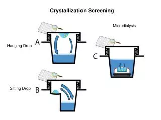
Practical No. 4 Hanging Drop Technique. Department of Microbiology College of Medicine. The Hanging-Drop Preparation ;

E N D
Practical No. 4 Hanging Drop Technique Department of Microbiology College of Medicine
The Hanging-Drop Preparation; • Since you have been oriented to some basic tools and methods used in microbiology, we shall begin our study of microorganisms by learning how to prepare them for study of their morphology under the microscope. • The simplest method for examining living microorganisms is to suspend them in a fluid (water, saline, or broth) and prepare a ''hanging drop,'' using a cover glass and a hollow ground slide. The slide is ground with a concave well in the center; the cover glass holds a drop of the suspension. • When the cover glass is inverted over the well of the slide, the drop hangs from the glass in the hollow concavity of the slide. Microscopic study of such a wet preparation can provide very useful information. Primarily the method is used to determine whether an organism is motile or not. Such a slide can be observed for a fairly long time period, because the drop does not dry up quickly.
Objective : To observe bacteria in a wet mount and determine their motility. Materials 24-hour broth culture of Proteus vulgaris 24-hour broth culture of Staphylococcus epidermidis. 2 hollow-ground slide Several cover glasses Wire inoculating loop Bunsen burner China-marking pencil Petroleum jelly
Site of flagella Peritrichous (E.coli) Monotrichous ( Vibrio cholerae)
Lophotrichous (pseudomonas) amphitrichous (Spirillum volutans) Site of flagella
Types of motility: 1-True movement (depends on flagella) 2- False movement. A- Brownian movement - Vibratory movement B- Drafting movement - organisms streaming along a tide
Three ways for detecting motility: 1- Flagella staining. 2- Motility test in semi-solid media. 3- Hanging drop technique.
1- Flagella staining Rosanalin dyesilver nitrate + ferric tannate
2. Semi-Solid media Inoculation The most commonly used test for motility in microbiology lab. It depends on the ability of motile bacteria to move through semi-solid media. Ordinary solid media contain 1.5-2.0% Agar Semi solid media contain about 0.4% Agar
Procedure of Motility Test • How to Perform Test: • Using a sterile bacteriological needle, pick a colony of the test organism • Stab quickly a tube of semi solid media. (avoid using bent needles). • Incubate the semi solid media for 24 hours
Reading Results: If bacteria is motile, there will be growth going out away from the stab line, and test is positive. If bacteria is not motile, there will only be growth along the stab line. A colored indicator can be used to make the results easier to see.
Procedures • 1-Take a cover glass and clean it thoroughly, making certain it is free of grease (the drop to be placed on it will not hang from a greasy surface). It may be dipped in alcohol and polished dry with tissue; or washed in soap and water, rinsed completely, and wiped dry. • 2-Take one hollow-ground slide and clean the well with a piece of dry tissue. Place a film of petroleum jelly around the rim of the well. • 3- Gently shake the broth culture of Proteus until it is evenly suspended. Using the wire inoculating loop, sterilize on the Bunsen flame, remove a loopful of culture. Close, and return the tube to the rack. • 4- Place the loopful of culture in the center of the cover glass (do not spread it around). Flame the loop and put it down. • 5- Hold the hollow-ground slide inverted, well down, over the cover glass; then press it down lightly so that the petroleum jelly adheres to the four edges of the cover glass. Now turn the slide over. You should have a sealed wet mount, the drop of culture hanging in the well.
6- Place the slide on the microscope stage, cover glass up. Start your examination with the low-power objective to find the focus. It is helpful to focus first on one edge of the drop, which will appear as a dark line. • The light should be reduced with the iris diaphragm, and, if necessary, by lowering the condenser. If you have trouble with the focus, ask the instructor for help. • 7- Continue your examination with the high-dry and oil immersion objectives (be very careful not to break the cover slip with the latter). • 8- Make a hanging-drop preparation of the staphylococcus culture, following the same procedure described above. • 9- Record your observation of the shape, cell groupings, and motility of the organisms. • 10- Discard your slides in a container with disinfectant solution.
Note: True independent motility of bacteria depends on their possession of flagella. If so equipped, they can propel themselves with progressive, directional locomotion (often quit rapidly). This kind of active motion must be distinguished from the vibratory movement of organisms or other particles suspended in the fluid. The latter type of motion is caused by the continuous, rapid oscillation of molecules of the fluid. • Small particles of any kind, including bacteria (whether motile or not), are constantly bombarded by the vibration of the fluid molecules, and so are bobbed up and down, back and forth. Such movement is irregular and non-directional and does not cause non motile organisms to change position with respect to other objects around them. • One must be careful not to mistake movement caused by currents in a liquid for true motility. If a wet mount is not well sealed or contains bubbles, air currents set up reaction fluid currents, and one sees organisms streaming along on a tide.
Colonial Morphology; • Some bacteria divide every 10 minutes, others may be every 30 minutes, 1 hour, or 2 hours, in case of Mycobacterium tuberculosis ( the causative agent of tuberculosis) may divide every 8-12 hours, while Mycobacterium leprae ( the causative agent of leprosy) divide once every 2 weeks. • In any of the above mentioned types, every single bacterium will divide repeatedly with time forming an aggregate of billions of cells which is called a colony. Every type of bacteria has its specific colonial characteristics that is very useful and important in diagnosis and differentiation. Such colonial characteristics are; • Color; some bacteria may produce pigments that colors the colony only without coloring the surroundings, and it is called non diffusible pigment. The other type of pigment produced by bacteria is that which is produced extracellularly and diffused in the surrounding medium leading to its coloration, and this type is called diffusible pigment.
Consistency; this character depends on the ability of the bacteria to produce large capsule, small capsule, slime or do not produce any. Therefore it makes the colony either; viscous, slightly viscous, or even watery. This character is tested by using a sterile needle and picks the tip of the colony and observe if a thread of viscous material is formed or not. • Surface texture; the colonial surface may be either; smooth, rough, granular, radiated….etc. • Shape; could be regular irregular, lobulated, radiated…etc. • Elevation; could be flat, slightly elevated, dome-shape or umbonate. • Margin; Either smooth, crenated, undulated or irregular. • Size; This is by measuring the diameter of the colony using a small ruler. • Odor; Some organisms produce colonies with a very distinctive odor that could be very helpful in identification, such as that produced by Proteus, Pseudomonas, and yeasts….etc. • Exercise; from the different types of colonies supplied by the instructors, try to write down and draw the different characters of the colonies you see with the help of a magnifier.