Download
1 / 18
180 likes | 305 Views
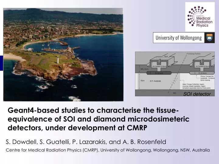
S. Dowdell, S. Guatelli, P. Lazarakis, and A. B. Rosenfeld Centre for Medical Radiation Physics (CMRP), University of Wollongong, Wollongong, NSW, Australia. Geant4-based studies to characterise the tissue-equivalence of SOI and diamond microdosimeter ic detectors, under development at CMRP.

E N D
S. Dowdell, S. Guatelli, P. Lazarakis, and A. B. Rosenfeld Centre for Medical Radiation Physics (CMRP), University of Wollongong, Wollongong, NSW, Australia Geant4-based studies to characterise the tissue-equivalence of SOI and diamond microdosimeteric detectors, under development at CMRP SOI detector
Silicon microdosimetry Microdosimetry measures energy deposition on a cellular scale Silicon On Insulator (SOI) microdosimeters have been under investigation at CMRP for the past ten years as a possible alternative to tissue equivalent gas counters Applications in radiation protection in space and dosimetry in medical physics Collaboration of CMRP with Australian Nuclear Science and Technology Organisation University of New South Wales, Australia First generation SOI detector Second generation SOI detector
Geant4 for microdosimetry • Characterisation of SOI microdosimeter • Response of the detector in radiation fields of interest • Proton therapy • Fast neutron therapy • Radiation protection in terrestrial, aviation and space • Chord length distribution • Tissue equivalence • Methodology to convert microdosimetric spectra in silicon to tissue
Geant4 for microdosimetry • Advanced capability in physics and geometry modelling • Good agreement between experimental and simulated microdosimetric spectra A. B. Rosenfeld, A. J. Wroe, I. M. Cornelius, M. Reinhard, and D. Alexiev, “Analysis of inelastic interactions for therapeutic proton beams using Monte Carlo simulation”, IEEE Trans. Nucl. Sci, vol. 51, no. 6, pp. 3019-3025, 2004. A. B. Rosenfeld, A. Wroe, M. Carolan, and I. Cornelius, “Method of Monte Carlo simulation verification in hadrontherapy with non-tissue equivalent detectors”, Rad. Prot. Dosim., vol. 119, pp. 487-490, 2006. D. A. Prokopovich, M. I. Reinhard, I. M. Cornelius, and A. B. Rosenfeld, “SOI microdosimetry for mixed field radiation protection”, Rad. Meas., vol. 43, pp. 1054-1058, 2008.
Characterisation of SOI microdosimetersby means of Geant4 Chord length distribution Tissue-equivalence of silicon
R1 = 3µm H = 2µm R2 Chord Length Distribution study The detector concept is exposed to a uniform isotropic field of infinite straight lines 2 µm device 10 µm device Mean chord length: (2.103 ± 0.014) µm Mean chord length: (3.36 ± 0.02) µm Geant4 Log scale Log scale Geant4
Characterisation of SOI microdosimetersby means of Geant4 Chord length distribution Tissue-equivalence of silicon S. Guatelli, et al., “Tissue equivalence correction in silicon microdosimetry for protons characteristic of the LEO space environment”, IEEE Trans. Nucl. Sci., vol. 55, pp. 3407-3413, 2008
Geant4 simulationHow to convert Si based microdosimetric measurements to tissue Experimental set-up Blue tracks: proton tracks Red tracks: e- tracks • Microdosimetric energy deposited spectra along the proton Bragg peak • in a simplified model of the Si SV • in a cylinder of water • Experimental set-up • 50, 100, 150, 200 MeV proton pencil beam • Electromagnetic and hadronic physics active for primary and secondary particles • Calculate the energy deposit per event in the detector Water phantom (size= 10 m) 10 μm 5 μm D Si detector
Geant4 simulation: Physics List • Electromagnetic physics: G4 Standard Package • Threshold of production of secondary particles: 1 micrometer • Hadronic physics for protons, neutrons and pions: • Geant4 elastic process • Parameterised Elastic Model • Inelastic hadronic processes • Bertini cascade model • Fission and capture processes • Inelastic hadronic scattering for deuteron, triton and α particle • Low Energy Parameterized model • The Binary Ion Model was active in the case of other ions
Proton Bragg peak in water 50 MeV 100 MeV 250 MeV 150 MeV The result of the study is the factor C, corresponding to the best agreement of the energy deposition spectra in the Si and in the water microdosimeter sites. For a water cylinder with height h and diameter d , the required scaling is C∙h, C∙d of silicon.
Results Statistical analysis by means of Kolmogorov-Smirnov test Energy deposition spectra resulting from a 50 MeV at 2 cm depth in the water phantom. (raising part of Bragg curve) • Linear coefficient C* = (0.56 ± 0.03) was determined to convert microdosimetric spectra from silicon to water for incident proton energies between a few MeV and 250 MeV • For a water cylinder with height h and diameter d, the required scaling is C*·h, C*·d of silicon Energy deposition spectraresulting from a 50 MeV proton beam at 2.1 cm depth in the water phantom. (Bragg peak)
Work in progress • We are investigating a general methodology to convert silicon microdosimetric spectra to tissue equivalent spectra in any arbitrary mixed radiation field • Study of the tissue equivalency of diamond • Diamond proposed as alternative to silicon for microdosimetry because of better tissue-equivalency
Secondary neutrons in proton therapy Secondary neutrons are generated through nuclear interactions of the primary beam with the beamline and inside the patient Neutrons deposit energy through secondary charged particles, often of high LET, and are important for second cancer calculations Accurate determination of secondary neutron dose delivered in tissue is vital Using different phantoms may lead to different secondary neutron doses Geant4-based study of secondary neutron dose in different materials S. Dowdell, et al., “Tissue equivalence of phantom materials for neutron dosimetry in proton therapy”, Med Phys, accepted for publication The materials considered were: A150, brain, cranium, lucite, muscle, water
Geant4 simulations • Neutron absorbed dose scored in sensitive volumes (2.5 x 25.0 x 0.5 mm3)throughout different materials • Absorbed dose (D) combined with radiation weighting factor (wR) to give dose equivalent H • H = wR x D 150 MeV p beam (5 cm x 5cm) • Physics list based on previous study by Zacharatou Jarlskog and Paganetti*: • G4EmStandard • G4BinaryCascade • G4UHadronElasticProcess • Cut in material = 1mm • Cuts in sensitive volume = 1mm * C. Zacharatou Jarlskog, and H. Paganetti, "Physics Settings for Using the Geant4 Toolkit in Proton Therapy", IEEE Trans. Nucl. Sci., 55, 1018-1025, (2008) Simulation setup
affecting out of field measurements where neutrons dominate the total dose equivalent Results • Magnitude of secondary neutrons doses out of field is less than along central axis, but larger differences exist between phantom and tissue • Secondary neutron dose in proton therapy is sensitive to the materials traversed Neutron absorbed dose (a) and dose equivalent (b) along the central axis
Next step: in progress How different are microdosimetric spectra obtained by silicon microdosimeter in different phantom materials irradiated with the same proton beam? How similar are phantom materials to tissue of interest for modelling of radiobiological properties of the proton beam?
Geant4 simulation experimental setup • 150 MeV and 250 MeV proton beams simulated • Energy deposition spectra collected in Si sensitive volume (red sphere), at multiple depths in different phantoms and tissues • The energy deposition spectra will be converted to microdosimetry spectra in Si Proton pencil beam • Significance of any variation in the spectra will be ascertained using the Kolmogorov-Smirnov statistical test • This study will show any effect that the phantom material has on a microdosimetric level Si Phantom R1 = 10m R2 = 3mm R3 = 10mm Geant4 simulation experimental setup
Summary At the CMRP we are intensively using Geant4 as a simulation toolkit to characterise novel detectors in radiation fields of interest for medical physics and radiation protection Thank you