Download
1 / 61
650 likes | 1.06k Views
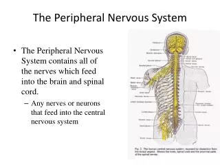
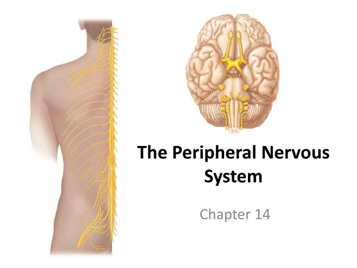
The Peripheral Nervous System. Chapter 14. The Peripheral Nervous System (PNS). The nervous system outside the brain and spinal cord (CNS) Provides vital links to the body and outside world Nerves allow the CNS to receive information and initiate action

E N D
The Peripheral Nervous System Chapter 14
The Peripheral Nervous System (PNS) • The nervous system outside the brain and spinal cord (CNS) • Provides vital links to the body and outside world • Nerves allow the CNS to receive information and initiate action • Sensory inputs and motor outputs categorized as • Somatic or visceral • General or special • Autonomic nervous system (ANS): general visceral motor part of the PNS • Two divisions: parasympathetic and sympathetic
Central nervous system (CNS) Peripheral nervous system (PNS) Sensory (afferent) division Motor (efferent) division Somatic sensory General: Touch, pain, pressure, vibration, temperature, and proprioception in skin, body wall, and limbs Visceral sensory General: Stretch, pain, temperature, chemical changes, and irritation in viscera; nausea and hunger Somatic nervous system Autonomic nervous system (ANS) Motor innervation of all skeletal muscles Motor innervation of smooth muscle, cardiac muscle, and glands Special: Hearing, equilibrium, vision Special: Taste, smell Sympathetic division Parasympathetic division Functional Organization of the PNS Figure 14.1
Basic Structural Components of the PNS • Sensory receptors: pick up stimuli from inside or outside the body • Nerves: bundles of peripheral axons • Ganglia: clusters of peripheral neuronal cell bodies • Motor endings: axon terminals of motor neurons • Innervate effectors (muscle fibers and glands)
Peripheral Sensory Receptors • Structures that pick up sensory stimuli that initiate signals in sensory axons • General sensory receptors – widely distributed nerve endings of sensory neurons monitor: • Touch, Pressure, Vibration, Stretch, Pain, Temperature, Proprioception • Two main categories of sensory receptors • Free nerve endings of sensory neurons: monitor general sensory information • Complete receptor cells: specialized epithelial cells or small neurons that monitor most types of special sensory information • Sensory receptors also classified according to • Location, type of stimulus detected, and structure
Classification by Location • Exteroceptors: sensitive to stimuli arising from outside the body • Located at or near body surfaces • Include receptors for touch, pressure, pain, and temperature • Interoceptorsreceive stimuli from internal viscera • Located in digestive tube, bladder, and lungs • Monitor a variety of stimuli: changes in chemical concentration, taste stimuli, stretching of tissues, temperature • Proprioceptors located in skeletal muscles, tendons, joints, and ligaments • Monitor degree of stretch • Send inputs on body movement to the CNS
Classification by Stimulus Detected • Mechanoreceptors: respond to mechanical forces • Touch, pressure, stretch, vibration, and itch • Baroreceptors monitor blood pressure • Thermoreceptors: respond to temperature changes • Chemoreceptors: respond to chemicals in solution • Photoreceptors: respond to light, located in the eye • Nociceptors: respond to harmful stimuli that result in pain
Classification by Structure • General sensory receptors divided into two groups: • Free nerve endings: abundant in epithelia and underlying connective tissue • Respond to pain and temperature • Monitor affective senses • Encapsulated nerve endings: consist of one or more end fibers of sensory neurons • Enclosed in connective tissue
Structure of free and encapsulated general sensory receptors Tactile corpuscle (touch, light pressure) Epithelial tactile complexes (light touch) Free nerve endings (pain and temperature) Epidermis Dermis and hypodermis Lamellar corpuscle (deep pressure) Hair follicle receptor (hair movement) Bulbous corpuscle (pressure)
Free Nerve Endings • Two specialized types of free nerve endings • Epithelial tactile complexes (Merkel discs): consist of tactile epithelial cell innervated by sensory nerve ending • Slowly adapting receptors for light touch • Hair follicle receptors: wrap around hair follicles, rapidly adapting receptors
Unencapsulated Nerve Endings Table 14.1 (1 of 4)
Encapsulated Nerve Endings • Mechanoreceptors include four main types • Tactile (Meissner’s) corpuscles • Lamellar (Pacinian) corpuscles • Bulbous corpuscles (Ruffini endings) • Proprioceptors
Tactile Corpuscles • Tactile (Meissner’s) corpuscles • Spiraling nerve ending surrounded by Schwann cells • Occur in the dermal papillae • Rapidly adapting receptors for discriminative touch • Occur in sensitive, hairless areas of the skin Table 14.1 (2 of 4)
Encapsulated Nerve Endings • Lamellar Corpuscles: single nerve ending surrounded by layers of flattened Schwann cells • Occur in the hypodermis • Sensitive to deep pressure—rapidly adapting receptors • Bulbous Corpuscles: located in the dermis and respond to pressure • Monitor continuous pressure on the skin—adapt slowly • Proprioceptors: monitor stretch in locomotory organs • Three types of proprioceptors
Lamellar Corpuscles and Bulbous Corpuscles Table 14.1 (3 of 4)
Three Types of Proprioceptors • Muscle spindles: measure the changing length of a muscle • Imbedded in the perimysium between muscle fascicles • Golgi tendon organs: located near the muscle-tendon junction • Monitor tension within tendons • Joint kinesthetic receptors: sensory nerve endings within the joint capsules
Secondary sensory endings (type II fiber) Efferent (motor) fiber to muscle spindle Efferent (motor) fiber to extrafusal muscle fibers Primary sensory endings (type Ia fiber) Extrafusal muscle fiber Muscle spindle Intrafusal muscle fibers Connective tissue capsule Sensory fiber Tendon organ Tendon Structure of Proprioceptors Figure 14.3
Proprioceptors Table 14.1 (4 of 4)
Cranial Nerves • Attach to the brain and pass through foramina of the skull • Numbered from I–XII • Cranial nerves I and II attach to the forebrain • All others attach to the brain stem • Primarily serve head and neck structures • The vagus nerve (X) is the only cranial nerve that extends into the abdomen
Filaments of olfactory nerve (I) Frontal lobe Olfactory bulb Olfactory tract Optic nerve (II) Temporal lobe Optic chiasma Optic tract Oculomotor nerve (III) Infundibulum Trochlear nerve (IV) Facial nerve (VII) Vestibulocochlear nerve (VIII) Trigeminal nerve (V) Abducens nerve (VI) Glossopharyngeal nerve (IX) Vagus nerve (X) Cerebellum Accessory nerve (XI) Medulla oblongata Hypoglossal nerve (XII) The Cranial Nerves Figure 14.4a
Cranial nerves Sensory function Motor function Somatic sensory (SS) Visceral sensory (VS) Somatic motor (SM) Visceral motor: parasympathetic (VM) I Olfactory Smell II Optic Vision III Oculomotor SM VM IV Trochlear SM General V Trigeminal SM VI Abducens SM (b) The Cranial Nerves Figure 14.4b (1 of 2)
Cranial nerves Sensory function Motor function Somatic sensory (SS) Visceral sensory (VS) Somatic motor (SM) Visceral motor: parasympathetic (VM) VII Facial General General; taste SM VM VIII Vestibulocochlear Hearing; equilibrium Some IX Glossopharyngeal General General; taste SM VM X Vagus General General; taste SM VM XI Accessory SM XII Hypoglossal SM (b) The Cranial Nerves Figure 14.4b (2 of 2)
Olfactory Nerves • Sensory nerves of smell Table 14.2 (1 of 12)
II The Optic Nerves • Sensory nerve of vision Table 14.2 (2 of 12)
III The Oculomotor Nerves • Innervates four of the extrinsic eye muscles Table 14.2 (3 of 12)
IV The Trochlear Nerves • Innervates the superior oblique muscle (an extrinsic eye muscle) Table 14.2 (4 of 12)
V The Trigeminal Nerves Table 14.2 (5 of 12)
The Trigeminal Nerves • Largest of the cranial nerves has three divisions • Ophthalmic division (V1) • Maxillary division (V2) • Mandibular division (V3) • Cell bodies of sensory neurons located in the trigeminal ganglion • Mandibular division contains motor fibers that innervate the chewing muscles
VI The Abducens Nerves • Abducts the eyeball—innervates lateral rectus muscle Table 14.2 (6 of 12)
VII The Facial Nerves • Innervates muscles of facial expression Table 14.2 (7 of 12)
VIII The Vestibulocochlear Nerves • Sensory nerve of hearing and balance Table 14.2 (8 of 12)
IX The Glossopharyngeal Nerves • Innervates structures of the tongue and pharynx Table 14.2 (9 of 12)
X The Vagus Nerves • A mixed sensory and motor nerve • “Wanders” into thorax and abdomen • Parasympathetic innervation of organs Table 14.2 (10 of 12)
XI The Accessory Nerves • Unique among cranial nerves • Accessory nerves are formed from ventral rootlets of the spinal cord • Do not arise from the brainstem Table 14.2 (11 of 12)
XII The Hypoglossal Nerves • Runs inferior to the tongue innervates the tongue muscles Table 14.2 (12 of 12)
Spinal Nerves • 31 pairs—contain thousands of nerve fibers • Connect to the spinal cord • Named for point of issue from the spinal cord • 8 pairs of cervical nerves (C1–C8) • 12 pairs of thoracic nerves (T1–T12) • 5 pairs of lumbar nerves (L1–L5) • 5 pairs of sacral nerves (S1–S5) • 1 pair of coccygeal nerves (Co1)
Cervical plexus Cervical nerves C1 – C8 Brachial plexus Cervical enlargement Thoracic nerves T1 – T12 Intercostal nerves Lumbar enlargement Lumbar nerves L1 – L5 Lumbar plexus Sacral nerves S1 – S5 Sacral plexus Coccygeal nerve Co1 Cauda equina Spinal Nerves Posterior View Figure 14.5
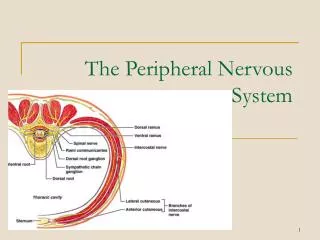
Spinal Nerves • Connect to the spinal cord by the dorsal root and ventral root • Dorsal root—contains sensory fibers • Cell bodies—located in the dorsal root ganglion • Ventral root—contains motor fibers arising from anterior gray column • Branch into dorsal ramus and ventral ramus • Dorsal and ventral rami contain sensory and motor fibers • Dorsal rami innervate back muscles follow a neat, segmented pattern • Innervate a horizontal strip of muscle and skin n iline with emergence point from the vertebral column • Rami communicantes connect to the base of the ventral ramus leading to the sympathetic chain ganglia
Sensory axon and cell body Dorsal root ganglion Dorsal root Dorsal ramus Nerves Spinal nerve Ventral ramus Ventral root Axon of motor neuron Neuromuscular junction Sensory receptors in skin (e.g., free nerve endings of sensory neuron) Spinal Nerves Figure 14.6
Spinal Nerves White matter Gray matter Ventral root Dorsal and ventral rootlets of spinal nerve Dorsal root Dorsal root ganglion Dorsal ramus of spinal nerve Ventral ramus of spinal nerve Spinal nerve Rami communicantes Sympathetic trunk ganglion (a) Anterior view showing spinal cord, associated nerves, and vertebrae. The dorsal and ventral roots arise medially as rootlets and join laterally to form the spinal nerve. Figure 14.7a
Dorsal ramus Ventral ramus Spinal nerve Rami communicantes Intercostal nerve Sympathetic trunk ganglion Dorsal root ganglion Dorsal root Ventral root Branches of intercostal nerve Lateral cutaneous Anterior cutaneous Sternum (b) Cross section of thorax showing the main roots and branches of a spinal nerve. Innervation of the Back
Innervation of the Anterior Thoracic and Abdominal Wall • Thoracic region • Ventral rami arranged in simple, segmented pattern • Intercostal nerves—supply intercostal muscles, skin, and abdominal wall • Each gives off lateral and anterior cutaneous branches
Introduction to Nerve Plexuses • Nerve plexus—a network of nerves • Ventral rami (except T2–T12) • Branch and join with one another • Form nerve plexuses • In cervical, brachial, lumbar, and sacral regions • Primarily serve the limbs • Fibers from ventral rami crisscross
Ventral rami Segmental branches Hypoglossal nerve (XII) Ventral rami: Lesser occipital nerve C1 Greater auricular nerve C2 Transverse cervical nerve C3 Ansa cervicalis C4 Accessory nerve (XI) C5 Phrenic nerve Supraclavicular nerves The Cervical Plexus • Buried deep in the neck under the sternocleidomastoid muscle • Formed by ventral rami of first four cervical nerves (cn 1–4) • Most are cutaneous nerves • Some innervate muscles of the anterior neck • Phrenic nerve—the most important nerve of the cervical plexus Figure 14.8
Major terminal branches (peripheral nerves) Roots (ventral rami) Cords Divisions Trunks Anterior Musculocutaneous C5 Upper Lateral Posterior Median C6 Medial Anterior Ulnar C7 Middle Posterior Radial C8 Posterior Anterior Lower Axillary T1 Posterior (c) Flowchart summarizing relationships within the brachial plexus The Brachial Plexus and Innervation of the Upper Limb • Brachial plexus lies in the neck and axilla • Formed by ventral rami of C5–C8 • Cords give rise to main nerves of the upper limb Figure 14.9c
Axillary nerve Humerus Radial nerve Musculocutaneous nerve Ulna Radius Ulnar nerve Median nerve Radial nerve (superficial branch) Dorsal branch of ulnar nerve Superficial branch of ulnar nerve Digital branch of ulnar nerve Muscular branch Median nerve (a) The major nerves of the upper limb Digital branch Nerves from the Lateral and Medial Cords • Musculocutaneous—main branch of the lateral cord • Innervates the biceps brachii and brachialis • Median—originates from both lateral and medial cords • Innervates anterior forearm muscles and lateral palm • Ulnar—branches from the medial cord • Innervates intrinsic hand muscles and skin of the medial hand Figure 14.9a
Roots (ventral rami) Anterior divisions C4 Dorsal scapular Posterior divisions C5 Nerve to subclavius Trunks C6 Suprascapular Roots Upper C7 Posterior divisions Trunks Middle C8 Lateral Lower T1 Cords Posterior Long thoracic Medial pectoral Medial Lateral pectoral Axillary Upper subscapular Musculo- cutaneous Lower subscapular Radial Thoracodorsal Median Medial cutaneous nerves of the arm and forearm Ulnar (b) Roots (rami C5–T1), trunks, divisions, and cords Nerves from the Lateral and Medial Cords Figure 14.9b
Nerves from the Lateral and Medial Cords Major terminal branches (peripheral nerves) Roots (ventral rami) Cords Divisions Trunks Anterior Musculocutaneous C5 Upper Lateral Posterior Median C6 Medial Anterior Ulnar C7 Middle Posterior Radial C8 Posterior Anterior Lower Axillary T1 Posterior (c) Flowchart summarizing relationships within the brachial plexus Figure 14.9c
Musculocutaneous nerve Axillary nerve Branches of axillary nerve Radial nerve Ulnar nerve (cut) Median nerve (cut) Posterior cutaneous nerve Deep radial nerve Superficial branch of radial nerve Anterior divisions Posterior divisions Nerves from the Posterior Cord • Radial—continuation of the posterior cord • Largest branch of the brachial plexus • Innervates muscles of the posterior upper limb • Axillary innervates the deltoid and teres minor Figure 14.12
Muscular innervation Cutaneous innervation Musculocutaneous nerve Median nerve Ulnar nerve C5 C5 C6 C6 C7 C7 C8 C8 T1 T1 Anterior view Posterior view Coraco- brachialis Biceps brachii Brachialis Medial cutaneous nerves off medial cord Pronator teres Flexor carpi radialis Flexor carpi ulnaris Palmaris longus Flexor digitorum superficialis Flexor pollicis longus Musculocutaneous nerve Flexor digitorum profundus Pronator quadratus Adductor pollicis 3 Thenar muscles Ulnar nerve 3 Hypothenar muscles 7 Interossei Lumbricals to digits 2,3 Lumbricals to digits 4,5 Median nerve Indicates variable contribution Axillary and Radial Nerves Figure 14.10