Download
1 / 29
340 likes | 582 Views
Abdominal trauma. Abdominal trauma. Any injury to the abdomen is called abdominal trauma, which can be either blunt or penetrating.

E N D
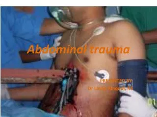
Abdominal trauma • Any injury to the abdomen is called abdominal trauma, which can be either blunt or penetrating. • In blunt trauma, clinical signs are not as evident, whereas in penetrating, abdominal trauma has obvious clinical signs, such as in stab wounds and gunshot wounds. • As it is with any penetration wound, there is increased risk of infection. Sometimes, it leads to damage to the organs found in the abdomen. • Abdominal wounds occur when there is a break in the continuity of the abdominal wall (either skin or mucous wall). • Signs and symptoms depend on the type of injury obtained. Usually, through ultrasonography, computed tomography and peritoneal lavage, a diagnosis can be made. Treatment will depend on the injury obtained aswell.
Signs & Symptoms • Past events of damage to the abdominal cavity • Pale, clammy skin • Evident injuries such as bleeding wounds, possibly with visible intestines • Severe pain possibly supplemented by muscle contractions across the abdominal wall • Vomiting and nausea • Contusions of the skin (bruising of the skin) • Incapable of standing up or and holding the injured area for pain relief • Incontinent (unable to control bladder or bowels) • Other hints of internal bleeding
Anatomy of the abdomen • The abdomen is enclosed by the abdominal cavity. • It is found below the ribcage and above the pelvic cavity. No bones are present in this area, unlike the chest and pelvic cavities. So when there are no bones to protect the abdomen and any injury may cause serious damage to some of the abdominal organs. • Some of the organs found in this region are from the different systems of the body. These include: • Integumentary system • Spleen • Urinarytract • Ureter • Kidneys Digestivetract • Stomach • Small intestine • Largeintestine (colon) • Liver • Gallbladder • Pancreas
Anatomy of the abdomen • The major organs of the abdomen include the small intestine, large intestine, and stomach. Because together, these three turn nutrients into usable energy, as well as helping to the disposal of solid waste. • Major organs that help filter contaminants out of the body are also in the abdominal region. These organs are the liver and kidneys. • The liver is located in the upper right-hand part of the abdominal cavity, just below the ribs. Although it has many functions, the liver is best known for processing blood, separating waste from nutrients. • Most people have two kidneys, although you can manage with just the one, which are located near the back of the body, under the ribs, on each side of the spine. Kidneys filter waste out of the bloodstream, which is passed out of the body as urine. The kidneys also helps to regulate levels of electrolytes, like salt and potassium and to produce certain hormones that play various roles throughout the body. • On top of the kidneys are the suprarenal (adrenal) glands. These synthesize and secrete hormones that help the kidneys to conserve sodium, thus conserving water. • The gallbladder is a tiny sack under the liver that holds extra bile made by the liver until it is pumped into the small intestine, which helps to break down fat. And the pancreas is yet another gland that produces enzymes to help your body digest proteins, carbohydrates, and fats. It also makes hormones that help regulate the distribution of nutrients, including sugar.
Muscles of the abdomen The abdominal muscles are located between the ribs and the pelvis on the front of the body. The abdominal muscles support the trunk, allow movement and hold organs in place and also by regulating internal abdominal pressure (sneezing, coughing, lifting, childbirth). • Transversus abdominis – Is a muscle layer of the anterior and lateral abdominal wall. Its also the deepest muscle layer. It arises from the lateral third of inguinal ligament, from the anterior 3/4 of the inner lip of the iliac crest, from the inner surfaces of the cartilages of lower six ribs and from the lumbodorsal fascia. The anterior ending is as a aponeurosis (Spigelian fascia), whereas the lower endings, toghether with the internal oblique muscles, are inserted at the pubis and pectineal line forming a co-joint tendon called aponeurotic falx. Innervated by lower intercostal nerves as well as the iliohypogastric nerve and ilioinguinal nerve. Its main roles are to stabilise the trunk and be able to maintain the internal abdominal pressure. • Rectus abdominis – The rectus abdominis is a long strap muscle that extends the entire length of the anterior abdominal wall. It is broader above and lies close to the midline, being separated from it's fellow by the linea alba. When contracting, this muscle has the characteristic bulges that are called ‘the six pack’. The main function of the rectus abdominis is to move the body between the ribcage and the pelvis. It arises by two heads from the anterior aspect of the symphysis pubis and the pubic crest and it inserts into the 5th , 6th and 7th costal cartilages and the xiphoid process.The rectus abdominis is an important postural muscle but it also plays an important role in forced expiration and in increasing intra-abdominal pressure.
Muscles of the abdomen • External oblique muscles – These are the largest and most superficial ones which are located on each side of the rectus abdominis. The external oblique muscles allow the trunk to twist, but to the opposite side of whichever external oblique is contracting. It arises from the external surface and inferior borders of the lower eight ribs. The fibres from the lowest ribs pass downwards, inserted into the anterior half of the iliac crest. The middle and upper fibres, directed inferiorly and anteriorly, end in an aponeurosis at the mid-clavicular line into the xiphoid process, the linea alba, the pubic crest and the pubic tubercle. • Internal oblique muscles – these flank the rectus abdominis and are located just inside the hipbones. They operate in the opposite way to the external oblique muscles. For example, twisting the trunk to the left requires the left side internal oblique and the right side external oblique to contract together. It arises from the thoraco-lumbar fascia, the anterior two-thirds of the iliac crest and the lateral two-thirds of the inguinal ligament. The muscle fibres radiate superomedially and insert into the inferior borders of the lower three ribs and their costal cartilages, the xiphoid process, the linea alba and the symphysis pubis.
Surgical landmark - pyramidalis muscle • A small, triangular muscle which lies anterior to the rectus abdominis on both sides. • Contained in the rectus sheet, which is formed by the aponeurosis of the transversus abdominis, external and internal oblique muscles. • Absent in 20% of the human population, either on one side or both sides. If absent —> then the lower end of the rectus becomes increased in size. • It can also be used as a surgical landmark —> while making the longitudinal incision for a classical caesarean section, the pyramidal muscle is use to determine midline and the location of linea alba.
Polytrauma • Injury to several (>1) physical regions or organ systems, where at least one injury or the combination of several injuries is life threatening with the severity of injury being > 16 on the scale of the Injury Severity Score (ISS) • The Injury Severity Score (ISS) is an anatomical scoring system that provides an overall score for patients with multiple injuries. Each injury is assigned an Abbreviated Injury Scale (AIS) score and is allocated to one of six body regions (Head, Face, Chest, Abdomen, Extremities (including Pelvis), External). Only the highest AIS score in each body region is used. The 3 most severely injured body regions have their score squared and added together to produce the ISS score. • To be differentiated from multiple injuries, which is not life threatening, or a severe, life-threatening single injury (barytrauma)
Causes • Road traffic accidents • Industrial accidents • Sport accidents • Accidents in leisure time • Accidents in the home • Violent crimes • Burials • Suicide attempts • Catastrophes • Effects of war • Accidents caused by internal conditions (e.g. heart attack)
First aid - abdominal wounds/trauma • Position and assist the patient in a position most comfortable to him/ her. This is usually on the back or the uninjured side. Flex the knees (drawn up) to relieve of pain and spasm. • Any tight clothing should be relieved, especially at the neck and waist. Sustain the patient with pillows and blankets for comfort and to avoid loss of body heat, respectively. Calm and reassure the patient. • Call for paramedics. • Assess the damage. Find entry and exit wounds. To check the back of the victim, use the hand to feel for a wound and find a pool of blood. If there is more than one open abdominal wound, the more serious wound should be treated. 5. Remove, cut or tear the clothing to expose the open abdominal wound. If clothing is stuck to the wound, cut or tear around the stuck clothing. Foreign objects on the wound must not be removed. In chemical environments, do not expose the wound. 6. If part of any organs has been dislodged, do not attempt to put it back. Using a sterile, dry material, gradually lift the organ and place on top of the victim’s abdomen. 7. Cover the wound using a sterile dressing. If it is a protruding wound, stabilize the object using a clean material and bandage. Using one hand, keep the dressing in place to prevent slipping.
Imaging options • Plain X-ray -plays a limited role. Used to evaluate presence of thoracic or pelvic injuries. Common findings include pneumothorax and hemothorax. In the case of gunshot wounds, x-rays identify the location and number of retained projectiles. • Ultrasound - Initial assessment of blunt abdominal trauma. Serves as a screening function because it assesses for the presence of free fluid in the abdomen or pericardium. • Computed tomography - For stable patients.he information provided by CT allows prognosis of injury and selective nonoperative management in both blunt and penetrating trauma. It assesses solid organ hematomas and sentinel hematoma. • DPL • FAST method
Diagnostic Peritoneal Lavage = DPL • Is a invasive surgical diagnostic procedure to determine if there is any free floating fluid (mostly blood) in the abdominal cavity. • At one time, diagnostic peritoneal lavage (DPL), it was the diagnostic test of choice to detect bleeding within the abdominal cavity after trauma. Now it is replaced by CT and FAST. • Limited to the diagnosis of intra-abdominal hemorrhage in unstable trauma patients.
DPL- procedure • Application of locl anesthesia • Vertical skin incision is made, 1/3 of the distance from the umbilicus to the pubic symphysis. • The line alba is divided and the peritoneum entered after it has been picked up to prevent bowel perforation. • A catheter is inserted towards the pelvis and aspiration of material attempted using a syringe. If no blood is aspirated, 1 litre of warm 0.9% saline is infused and after a few minutes this is drained and sent for analysis.
Interpretation of results • If any of these are found then the DPL is (+) and the patient should be send to surgery: • RBC > 100.000mm3 • WBC > 500mm3 • Amylase levels > 175 IU/ml • Bilirubin level > 0.01 mg/DL • Alkaline phosphate levels ≥ 3 IU/L
FAST method • Focused Assessment with Sonography for Trauma (FAST) is a limited ultrasound examination directed solely at identifying the presence of free intraperitoneal or pericardial fluid. • FAST is less invasive than DPL, involves no exposure to radiation and is cheaper compared to CT, but achieves a similar accuracy • FAST examines four areas for free fluid: 1 Perihepatic & hepato-renal space (also called Morison’s pouch or hepatorenal recess) 2 Perisplenic space 3 Pelvic space 4 Pericardium
Benefits of FAST • Decreases the time to diagnosis for acute abdominal injury in BAT • Helps accurately diagnose hemoperitoneum • Helps assess the degree of hemoperitoneum in BAT • Is noninvasive • Can be integrated into the primary or secondary survey and can be performed quickly, without removing patients from the clinical arena • Can be repeated for serial examinations • Is safe in pregnant patients and children, as it requires less radiation than CT • Leads to fewer DPLs; in the proper clinical setting, can lead to fewer CT scans (patients admitted to the trauma service and to receive serial abdominal examinations)
Interpretation of results • FAST is most useful in trauma patients who are hemodynamically unstable. • A positive FAST result is defined as the appearance of a dark ("anechoic") strip in the dependent areas of the peritoneum. • In the right upper quadrant this typically appears in Morison’s pouch (between the liver and kidney). This location is most useful as it is the place where fluid will collect with a supine patient. • In the left upper quadrant, blood may collect anywhere around the spleen (perisplenic space). • In the pelvis, blood generally pools behind the bladder (in the rectovesicular space). A positive result suggests hemoperitoneum; often CT scan will be performed if the patient is stable[13] or a laparotomy if unstable. In those with a negative FAST result, a search for extra-abdominal sources of bleeding may still need to be performed.[13]
Liver injury • It is the most vulnerable abdominal organ to blunt injury because of its size and location • Also vulnerable to penetrating trauma • As a result of impact (for example, a motor vehicle crash) or penetrating trauma (such as a knife or gunshot wound) • The liver is injured in approximately 5% of all the people admitted to the hospital for trauma. • Its tissue is very delicate and it also has a large blood supply and capacity, so when injured it presents a serious risk of shock. • Can become lacerated or contused so that a hematoma may develop. Which could in turn lead to the leakage of bile, but this is usually without any serious consequences. • If severely injured, the liver may cause exsanguination (bleeding to death), requiring emergency surgery to stop the bleeding. • Diagnosis is based upon the use of computed tomography (CT) or ultrasonography to detect liver injuries. Sometimes surgery is needed to determine the extent of the injury and to stop the bleeding.
Symptoms & Treatments • People with liver injury and severe bleeding have symptoms of shock • rapid heart rate • rapid breathing • cold, clammy, pale or bluish skin • People also have abdominal pain and tenderness because blood in the abdomen irritates the abdominal tissue. • When bleeding is severe, the abdomen may also be swollen. • Sometimes liver injuries heal without treatment. However, people must be hospitalized and watched closely to ensure that bleeding does not worsen. Sometimes blood transfusions may help. If the bleeding worsens or does not stop fairly quickly, doctors often first try to seal off the bleeding vessels without surgery. To seal the vessels, doctors pass a thin plastic catheter into the blood vessels in the groin and then up to the liver. Then they inject substances to seal the vessels. If this procedure does not stop the bleeding, surgery is usually done. Also, if bleeding was very severe from the beginning, surgery is usually done as soon as possible because in such cases sealing off blood vessels without surgery is rarely effective.
Spleen injury • Because of the spleen’s position in the upper left side of the abdomen, a severe blow to the stomach area can damage the spleen, tearing its covering, the tissue inside, or both. The tears range from small ones that stop bleeding spontaneously or to very large ones that cause potentially fatal hemorrhage. • Sometimes a collection of blood (hematoma) forms under the covering of the spleen or deep within it. • The spleen is one of the most, if not the most, commonly injured organ in the abdomen as a result of motor vehicle crashes, falls from a considerable height and beatings. Sometimes other abdominal organs also get damaged. • Enlargement of the spleen (for example, due to Epstein-Barr virus causing infectious mononucleosis) makes the spleen more susceptible to injury. • When the spleen is injured, blood may be released into the abdomen. The amount of bleeding depends on the size of the injury. A hematoma of the spleen does not bleed into the abdomen at first but may rupture and bleed in the first few days after injury, although rupture sometimes does not occur for weeks or months. • Diagnosis is made by CT or ultrasonography
Symptoms & Treatments • An injured or ruptured spleen can make the abdomen painful and tender. • Blood in the abdomen acts as an irritant and causes pain. The pain is in the left side of the abdomen just below the rib cage. Sometimes the pain can even be felt in the left shoulder. • The abdominal muscles contract reflexively and feel rigid. If enough blood leaks out, blood pressure falls and people feel light-headed, have blurred vision and confusion, and lose consciousness (faint). • Splenectomy is performed. However, removing the spleen can cause later problems, which includes an increased susceptibility to infections. Nowadays, small and moderate-sized injuries to the spleen can heal without surgery, although blood transfusions are sometimes required and people must be treated in the hospital. When surgery is necessary, usually the entire spleen is removed (splenectomy), but sometimes surgeons are able to repair a small tear. Some people take antibiotics to prevent infections, particularly when they have another disorder (such as sickle cell disease or cancer) that increases the risk of developing life-threatening infections.