Download

1 / 36
500 likes | 780 Views
NANOFIBER TECHNOLOGY: DESIGNING THE NEXT GENERATION OF TISSUE ENGINEERING SCAFFOLDS. C.P. Barnes 1 , S.A. Sell 1 , E.D. Boland 1 , D.G. Simpson 2 , G.L. Bowlin 1 1 Department of Biomedical Engineering, 2 Department of Anatomy and Neurobiology Virginia Commonwealth University, Richmond, VA.

E N D
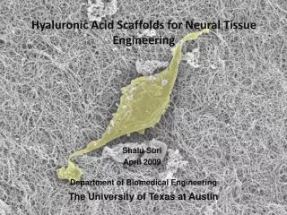
NANOFIBER TECHNOLOGY: DESIGNING THE NEXT GENERATION OF TISSUE ENGINEERING SCAFFOLDS C.P. Barnes1, S.A. Sell1, E.D. Boland1, D.G. Simpson2, G.L. Bowlin11Department of Biomedical Engineering, 2Department of Anatomy and NeurobiologyVirginia Commonwealth University, Richmond, VA MARK HWANG
Components - collagens - elastin - hyaluronic acid - proteoglycans - glycosaminoglycans - fibronectin EXTRACELLULAR MATRIX Signalling - cell adhesion - programmed cell death - migration - cytokine/growth factor activity - growth - differentiation
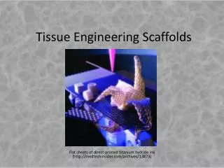
TISSUE ENGINEERING SCAFFOLDS - BACKGROUND Premise - ECM microenvironment key to tissue regeneration - Cell not viewed as self-contained unit Role of ECM - ECM mediates biochemical and mechanical signalling - Ideal ECM non-immunogenic promote growth maintain 3-D structure only native tissues remain post-treatment Research emphases to-date - Biocompatibility - Degradability
TISSUE ENGINEERING SCAFFOLDS - BACKGROUND Overall Goals - Design scaffold with maximum control over: biocompatibility (chemical) biodegradability (mechanical) - Utilize natural and synthetic polymers - Future directions: tissue regeneration drug delivery EFFECTIVE SCAFFOLD DESIGN BEGINS WITH ACCURATE SCALING Current Focus - Nanofiber synthesis
Scale difference necessary - single cell contacts thousands of fibers - transmission of fine/subtle signals NANOFIBERS - INTRODUCTION ECM fibers ~ 50-500 nm in diameter Cell ~ several-10 um Fibers 1-2 orders of magnitude < cell 3 techniques to achieve nanofiber scale - self assembly - phase separation - electrospinning
Example: peptide-amphiphiles - hydrophobic tail - cysteine residues disulfide bonds NANOFIBERS: SELF-ASSEMBLY • Definition: spontaneous organization into stable structure without covalent bonds • Biologically relevant processes • DNA, RNA, protein organization • - can achieve small diameter Drawbacks: more complex in vitro - limited to 1) several polymers and - 2) hydrophobic/philic interactions - small size; larger = unstable
NANOFIBERS: PHASE SEPARATION Definition: thermodynamic separation of polymer solution into polymer-rich/poor layers - similar to setting a gel - control over macroporous architecture using porogens, microbeads, salts 98% porosity achieved! - consistent Drawbacks: - limited to several polymers - small production scale
A- polymer solution in syringe B- metal needle C- voltage applied to need NANOFIBERS: ELECTROSPINNING Definition: electric field used to draw polymer stream out of solution D- electric field overcomes solution surface tension; polymer stream generated E- fibers 1) collected and 2) patterned on plate
NANOFIBERS: ELECTROSPINNING - simple equipment - multiple polymers can be combined at 1) monomer level 2) fiber level 3) scaffold level - control over fiber diameter alter concentration/viscosity - fiber length unlimited - control over scaffold architecture target plate geometry target plate rotational speed
NANOFIBERS: ELECTROSPINNING Drawbacks: - natural fibers 50-500 nm; spun fibers closer to 500 nm - architecture very random LACK OF GOLD STANDARD Current approaches combined techniques - usually electrospinning + phase separation - fibers woven over pores
ELECTROSPINNING POLYMERS Synthetics - Polyglycolic acid (PGA) - Polylactic acid (PLA) - PGA-PLA - Polydioxanone (PDO) - Polycaprolactone - PGA-polycaprolactone - PLA-polycaprolactone - Polydioxanone-polycaprolactone Natural - Elastin - Gelatin collagen - Fibrillar collagen - Collagen blends - Fibrinogen
POLYGLYCOLIC ACID (PGA) - biocompatible - consistent mechanical properties hydrophilic predictable bioabsorption (2-4 wks) - electrospinning yields diameters ~ 200 nm Parameters - surface area to volume ratio - spinning orientation affects scaffold elastic modulus Drawbacks - rapid hydrolitic degradation = pH change tissue must have buffering capacity
POLYGLYCOLIC ACID (PGA) Random fiber collection (L), aligned collection (R)
POLYGLYCOLIC ACID (PGA) Fiber collection Orientation affects stress / strain
POLYLACTIC ACID (PLA) – 200 nm - aliphatic polyester - L optical isomer used by-product of L isomer degradation = lactic acid - methyl group decreases hydrophilicity - predictable bioabsorption, slower than PGA (30 wks) - half-life ideal for drug delivery Parameters (similar to PGA) - surface area to volume ratio - spinning orientation affects scaffold elastic modulus Compare to PGA - low degradation rate = less pH change
POLYLACTIC ACID (PLA) – 200 nm Thickness controlled by electrospin solvent Chloroform solvent (L) ~ 10 um HFP (alcohol) solvent (R) ~ 780 nm Both fibers randomly collected
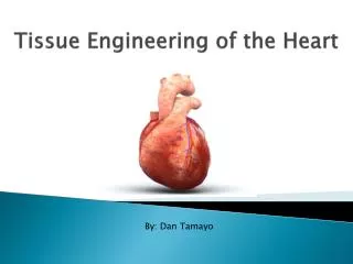
PGA+PLA = PLGA - tested composition at 25-75, 50-50, 75-25 ratios - degradation rate proportional to composition - hydrophilicity proportional to composition Recent Study - delivered PLGA scaffold cardiac tissue in mice - individual cardiomyocytes at seeding - full tissue (no scaffold) 35 weeks later - scaffold loaded with antibiotics for wound healing
PGA+PLA = PLGA PLGA modulus proportional to composition
POLYDIOXANONE (PDO) - crystalline (55%) - degradation rate between PGA/PLA close to 40-60 ratio - shape memory - modulus – 46 MPa; compare: collagen – 100 MPa elastin – 4 MPa Advantages - PDO ½ way between collagen/elastin, vascular ECM components - cardiac tissue replacement (like PLGA) - thin fibers (180nm) Drawbacks - shape memory – less likely to adapt with developing tissue
POLYCAPROLACTONE (PCL) - highly elastic - slow degradation rate (1-2 yrs) - > 1 um - similar stress capacity to PDO, higher elasticity Advantages - overall better for cardiac tissue – no shape retention bc elastic Previous Applications Loaded with: - collagen cardiac tissue replacement - calcium carbonate bone tissue strengthening - growth factors mesenchymal stem cell differentation
POLYCAPROLACTONE + PGA - PGA high stress tolerance - PCL high elasticity - optimized combination PGA/PCL ~ 3/1 - bioabsorption at least 3 mths (PCL-2 yrs, PGA 2-4 wks) Clinical Applications – none yet POLYCAPROLACTONE + PLA - PLA highly biocompatible (natural by products) - PCL high elasticity - more elastic than PGA/PCL - strain limit increases 8x with just 5% PCL
POLYCAPROLACTONE + PLA - PCL elastic; however, decreasing PLA/PCL ratios decreases strain capacity - strain capacity optimized at 95:5 - still ideal in vivo – mostly PLA = natural by products
POLYCAPROLACTONE + PLA Clinical Applications - several planned - all vasculature tissue - high PLA tensile strength react (constrict) to sudden pressure increase - increased elasticity with PCL passively accommodate large fluid flow OVERALL – passive expansion, controlled constriction = best synthetic ECM combination for cardiac application
POLYCAPROLACTONE + POLYDIOXANONE PCL PDO Recall… - PCL high elasticity - PDO approx = PLA/PGA - PDO shape memory – limits use in vascular tissue Findings - hybrid structure NOT = hybrid properties - lower tensile capacity than PDO - low elasticity than PDO - larger diameter - NOT clinically useful “[This] will be further investigated by our laboratory” In other words- not publishable, but 1 year’s worth of work and good enough for a master’s thesis
POLYCAPROLACTONE + POLYDIOXANONE PCL PDO Principle Drawbacks Large fiber diameter Low tensile/strain capacity Possible Cause? PDO is the only crystalline structure polymer
ELASTIN - highly elastic biosolid (benchmark for PDO) - hydrophobic - present in: vascular walls skin Synthesis of Biosolid? - 81 kDa recombinant protein (normal ~ 64 kDa) - repeated regions were involved in binding - 300 nm (not as small as PDO ~ 180 nm) - formed ribbons, not fibers – diameter varies Findings: - not as elastic as native elastin - currently combined with PDO to increase tensile strength - no clinical applications yet
COLLAGENS: FIBRIL FORMING Type I - 100 nm (not consistent) - almost identical to native collagen (TEM) - present is most tissues COLLAGENS: GELATIN - highly soluble, biodegradable (very rapid) - current emphasis on increasing lifespan Type II - 100-120 nm (consistent) - found in cartilage - pore size and fiber diameter easily controlled by dilution
Type II easy to regulate 1) fiber 2) pore size COLLAGENS: FIBRIL FORMING Type I (inconsistent fibers)
COLLAGENS: FIBRIL FORMING Type III - preliminary studies - appears consistent ~ 250 nm None of the electrospun collagens have clinical application yet
Scaffolds studied to-date - reconstructing the media: COLLAGENS BLENDS In context: vasculature - intima – collagen type IV + elastin - media – thickest, elastin, collagen I, III, SMC - adventia – collagen I
- cross section of tube wall - 5 day culture complete scaffold infiltration RECONSTRUCTING THE MEDIA - SMC seeded into tube - average fiber ~ 450 nm slightly larger ECM fibers - incorporation of GAG carbohydrate ECM collagen crosslinker mediate signalling
COMBINING COLLAGEN WITH PDO Observations: - collagen I highest tensile capacity - 70:30 collagen-PDO optimal ratio for all collagens
FIBRINOGEN - smallest diameter (both synthetic and bio) 80, 310, 700 nm fibers possible - high surface area to volume ratio increase surface interaction used in clot formation Stress capacity comparable to collagen (80-100 MPa)
HEMOGLOBIN - hemoglobin mats - clinical applications: drug delivery hemostatic bandages - fiber sizes 2-3 um - spun with fibrinogen for clotting/healing - high porosity = high oxygenation
OVERVIEW - Electrospinning viable for both synthetic and biological scaffolds/mats - Wide range of fiber sizes necessary and possible ECM ideally 150-500 nm cell mats 2-3 um - Hybridizing polymers can, but not necessarily, lead to hybrid properties Specifics: - PGA, PLA, PLGA most commonly used scaffold materials - PDO exhibits elastin+collagen functionality in 1 synthetic polymer BUT inhibited by “shape memory” - PCL most elastic synthetic – frequently mixed with other synthetics