Download
1 / 39
420 likes | 615 Views
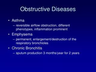
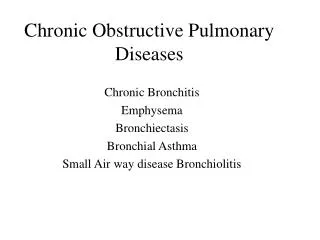
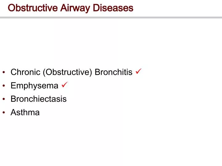
Obstructive Airway Diseases. Chronic (Obstructive) Bronchitis Emphysema Bronchiectasis Asthma . Bronchiectasis. Permanent bronchial dilation associated with suppuration (pus production) Occurs commonly with cystic fibrosis

E N D
Obstructive Airway Diseases • Chronic (Obstructive) Bronchitis • Emphysema • Bronchiectasis • Asthma
Bronchiectasis • Permanent bronchial dilation associated with suppuration (pus production) • Occurs commonly with cystic fibrosis • Results from infection and scarring (fibrous tissue formation) • Bronchi are dilated, due to ‘contraction’ effect of fibrous tissue • but can be fibrous obstruction leading to distal mucous retention and superimposed bacterial infection • Bronchi become filled with mucous and neutrophils, producing copious, foul smelling sputum • Secondary inflammation • further destruction of airways and alveolar walls • fibrosis of lung parenchyma
Asthma • Defined as increased irritability of the bronchial tree with paroxysmal airway narrowing • Accounts for more paediatric hospital admissions in UK than any other single cause • Two broad categories:- • Extrinsic (Allergic) • Intrinsic (Non-allergic) • Two types may co-exist
Asthma • Basis of intrinsic asthma is unknown • precipitated by non-allergenic factors • eg inhaled irritants, infection, stress, cold air, exercise • Basis of extrinsic asthma is immunologic • typically precipitated by type 1 (IgE-mediated) hypersensitivity reactions to inhaled allergens • immediate • but type 3 reactions may also be involved in some cases • triggered by soluble antigen/antibody immune complexes deposited in tissues • Variable presentation, from slight wheezing to severe dyspnoea
Asthma • Pathophysiology involves 3 main mechanisms:- • Reversible bronchoconstriction • Thickening of the bronchial mucosa • due to oedema and infiltration of inflammatory cells • Mucous plugging of the bronchiolar lumen • Allergic asthma has 2 phases • ‘early phase’ (occurring within minutes) • ‘late phase’ (occurring at 2-8 hours)
Allergic Asthma: Early Phase • Early phase consists of:- • Antigen exposure primes B lymphocytes • Consequent production of specific IgE • IgE binding to mast cells, eosinophils, macrophages and platelets • Release of inflammatory mediators, including histamine, various leukotrienes (LTB4, LTC4, LTD4, etc.) and prostanoids. • Processes occur within minutes • Hence known as the ‘early phase’ of an asthma attack
Allergic Asthma: Late Phase • Late phase consists of:- • Further release of inflammatory cytokines, including GM-CSF (Granulocyte Macrophage Colony-Stimulating Factor), IL4, IL5, TNF, etc. • These act as chemoattractants for eosinophils, neutrophils and other inflammatory cells • These, in turn, release leukotrienes, platelet activating factor (PAF), proteases, major basic protein (MBP), eosinophilic cation protein, etc. • These result in increased inflammation and oedema, mucous secretion, vasodilatation, bronchoconstriction and damage to the bronchial epithelium
Non-Allergic Asthma • Bronchial hypersensitivity may involve… • Increased mucosal inflammation • due to increased numbers of eosinophils? • Increased activity of irritant receptors and axon reflexes • release of bronchoconstrictor neuropeptides such as substance P ? • Increased vagal stimulation and decreased adrenergic stimulation of bronchial smooth muscle • bronchoconstriction and increased mediator release • Increased Ca2+ influx • into smooth muscle increased bronchoconstriction • into mast cells degranulation
Treatment of Obstructive Disorders • If possible treat the cause… • For COPD, this invariably means reducing exposure to noxious agents to limit (irreversible) damage • Reduce atmospheric pollution • Stop smoking! • Treat concurrent infections/allergies • It may be necessary, either additionally or alternatively, to treat the effects
Treatment of Obstructive Disorders • Treatment of cause may include:- • Antibiotics for infectious bacterial causes, or as prophylaxis against secondary bacterial infections in viral and other infections • Anti-allergic therapies in immune-induced disorders • Removal of physical obstructions • Treatment of effect may include:- • Bronchodilator agents • Anti-inflammatory agents • Others
Bronchodilator Agents • β2-Adrenoceptor agonists • Act primarily on bronchial smooth muscle β2-Adrenoceptors to increase intracellular cAMP • Additionally they may:- • Prevent mast cell degranulation • Decrease microvascular permeability, and hence oedema • Increase mucociliary transport • Primarily of short term use since prolonged use can lead to receptor down-regulation and reduced efficacy • which may be reversed with steroids • Main agents are • salbutamol, terbutaline, (short acting, inhaled) • salmeterol, eformoterol (long acting, used for prophylaxis)
Bronchodilator Agents • Muscarinic ACh antagonists • Ipratropium, oxitropium are examples • Administered by inhalation • Usually less effective bronchodilators than β2-Adrenoceptor agonists • Methylxanthines • Phosphodiesterase inhibitors • increase cAMP by reducing breakdown • Now usually only used as second line drugs • Not as specific as β2-agonists • Examples include theophylline and aminophylline
Anti-inflammatory agents • Glucocorticoids • No direct effect on bronchial calibre • Thought to work via inhibition of synthesis and release of prostanoids, leukotrienes and cytokines as well as on eosinophils • Also up-regulate β-receptors • Hydrocortisone (IV) and prednisolone (oral) are used for management of status asthmaticus • Progressive, unresponsive, severe asthma • Beclomethasone, Fluticasone, Budesonide, etc (inhalation) for more general use
Anti-inflammatory agents • Lipoxygenase inhibitors • Inhibits activity of 5-lipoxygenase • Main enzyme for synthesis of leukotrienes, eg LTB4, LTC4 and LTD4 • New group; first FDA-approved example is Zileuton • Others • Sodium chromoglycate and nedocromil sodium inhibit both early and late phases of asthma • Mode of action is unclear, but may involve • inhibition of mediator release from inflammatory cells, • suppression of axon reflexes • inhibition of action of Platelet Activating Factor (PAF)
Others Agents • Leukotriene receptor antagonists • block LTD4 receptors • inhibit inflammation, mucosal oedema and bronchoconstriction • Eg zafirlukast, montelukast • Histamine receptor antagonists • block H1 receptors • inhibit IgE-mediated neutrophil influx and PAF-mediated eosinophil influx • Eg terfenadine, astemizole, azelastine • Ca2+ channel antagonists • reduce Ca2+-dependent release of mucous and chemical mediators • Eg nifedipine, verapamil
Others Agents • K+ channel openers • hyperpolarises bronchial smooth musclebronchodilation • Egcromakalim • Antibodies • prevent the binding of IgE to mast cells • EgIgE MCAs • Nitric oxide (NO) donors/PAF antagonists • none yet available • NO as inflammatory marker • NO sensor as early warning for asthmatics?
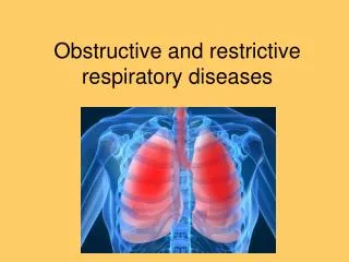
Restrictive Lung Disease • Already looked at Obstructive • Compromised airflow • Eg reduced FEV1 • Now briefly look at Restrictive • Reduced capacity • Eg reduced FVC
Acute Respiratory Distress Syndrome (ARDS) • Causes - Infection/sepsis/trauma/O2 toxicity • Risk Groups - Adults • Pathophysiology • Initiated by damage to alveolar endothelium & Type II pneumocytes • Impaired gas exchange due to pulmonary hemorrhage, pulmonary oedema, or atelectasis • Complement activation, sepsis • Features • Acute dyspnea, respiratory failure • Hypoxia (cyanosis) • Heavy, wet lungs
Sarcoidosis • Causes - Unknown • Risk Groups – Females, young, black • Pathophysiology • Small lumps (granulomas) due to chronic inflammation • Multi-system (eg lungs, lymph nodes, eyes, joints etc) • Features • Dyspnea on exertion • Dry cough, fever, fatigue • Uveitis, dry eyes • Polyarthritis
Hypersensitivity Pneumonitis (Farmer’s Lung) • Causes – Exposure to organic antigens • Risk Groups – Occupational (farms, birds, etc) • Pathophysiology • Type III & IV hypersensitivity reactions • Chronic interstitial inflammation • Alveolar damage fibrotic lung • Features • Acute or chronic • Dry cough • Chest tightness • General malaise, fever
Idiopathic Pulmonary Fibrosis • Causes – Unknown • Risk Groups – Male, 50-60 y, smoking • Pathophysiology • Chronic inflammation of alveolar wall fibrosis • Usually fatal within 4-6 years • Features • Velcro-like rales • Honeycomb lung (end stage) • Finger-clubbing • due to disorder in megakaryocyte fragmentation in lungs • aggregation in small vessels at finger tips?
TLC 9 8 7 VC VT TLC 6 VOLUME ERV 5 (litres) TLC 4 VC VT 3 VC VT RV 2 ERV RV 1 RV Restricted Normal COPD Lung Function in Obstructive/Restrictive Disease Restriction Normal Obstruction
Lung Volumes and Capacities • 4 Volumes:Inspiratory Reserve VolumeTidal VolumeExpiratory Reserve VolumeResidual Volume • 2 or more Volumes a Capacity • 4 Capacities:Vital CapacityInspiratory CapacityFunctional Residual CapacityTotal Lung Capacity
Spirometry • “Spirometry is a medical test that measures the volume of air an individual inhales or exhales as a function of time. (ATS, 1994)” • A spirometer can be used to measure the following: • FVC and its derivatives (such as FEV1, FEF 25-75%) • Forced Inspiratory Vital Capacity (FIVC) • Peak Expiratory Flow Rate (PEF) • Maximum Voluntary Ventilation (MVV) • Slow VC • IC, IRV and ERV • Pre- and post-bronchodilator tests
Flow-Volume Curves • Two ways to record/display results… • Volume as a function of time • Flow rate as a function of volume • (expiration & inhalation)
Measurement Definitions • FVC – the maximum volume exhaled • FEV1 – the volume exhaled during the first second of the FVC manoeuvre • FEV1% – the ratio of FEV1 to FVC expressed as a percentage • A reduction is specific for obstructive rather than restrictive diseases • FEF 25-75% – the mean expiratory flow rate during the middle half of an FVC manoeuvre • Reflects flow through the small (<2mm diameter) airways
Measurement Definitions • Estimate FEV1% and FEF 25-75%
Flow-Volume Loop • A plot of inspiratory/expiratory flow (y-axis) vs volume (x-axis) during maximal, forced manoeuvres
Obstructive Lung Disease - Asthma • PEF reduced (max height of the loop) • Airflow reduces rapidly post-PEF concave loop • Reversed with bronchodilator treatment • (Irreversible in COPD)
Obstructive Lung Disease - Emphysema • Airways collapse during forced expiration • Destruction of supporting lung tissue reduced flow at low lung volume • ‘dog-leg’ curve
Restrictive Lung Disease • Full lung expansion prevented by fibrotic tissue • Typically, reduced FVC (shown) • But PEF may be preserved/increased (not shown)
Obstructive vs. Restrictive Defect • Obstructive Disorders • FVC or↓ • FEV1 ↓ • FEF 25-75% ↓ • FEV1/FVC ↓ • TLC or ↑ • Restrictive Disorders • FVC ↓ • FEV1 ↓ • FEF 25-75% to ↓ • FEV1/FVC to ↑ • TLC ↓
Limits of Spirometry • Cannot measure TLC, FRC, RV
Body Plethysmography • Fully enclosed rigid box • Breathe through mouthpiece connected via shutter to the internal volume of the box • Breathe against the shutter • Pressure in lungs changes • Pressure in the box changes (opposite direction) • Application of Boyle’s Law lung volumes • Including parts of lungs not in communication with bronchial tree