Download
1 / 49
500 likes | 705 Views
Detecting Oral Cancer. What is cancer?. Cancer is a loss of growth regulation Cells grow when they shouldn’t tumour Cells grow where they shouldn’t invasion, metastasis. Cancer is a genetic disease of somatic cells.

E N D
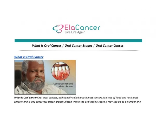
What is cancer? • Cancer is a loss of growth regulation • Cells grow when they shouldn’t tumour • Cells grow where they shouldn’t invasion, metastasis
Cancer is a genetic disease of somatic cells • Mutations in specific genes can cause a normal cell to become cancerous
What are these genes that lead to cancer when mutated? • Proto-Oncogenes • Gas pedal for cell proliferation • Mutation Oncogene Gas pedal stuck down • Tumour suppressor genes • Brakes for cell division • Mutation Brakes don’t work • Care taking (DNA repair) genes
Carcinogenesis • Carcinogensis is a multistep process involving mutation in multiple genes
Angiogenesis • Without blood supply tumour can grow 2mm (106 cells) • Produce growth factors to stimulate angiogenesis • Blood vessels around tumour is a bad sign!
Cancer is a loss of growth regulation induced by: • Environmental factors • Genetic factors
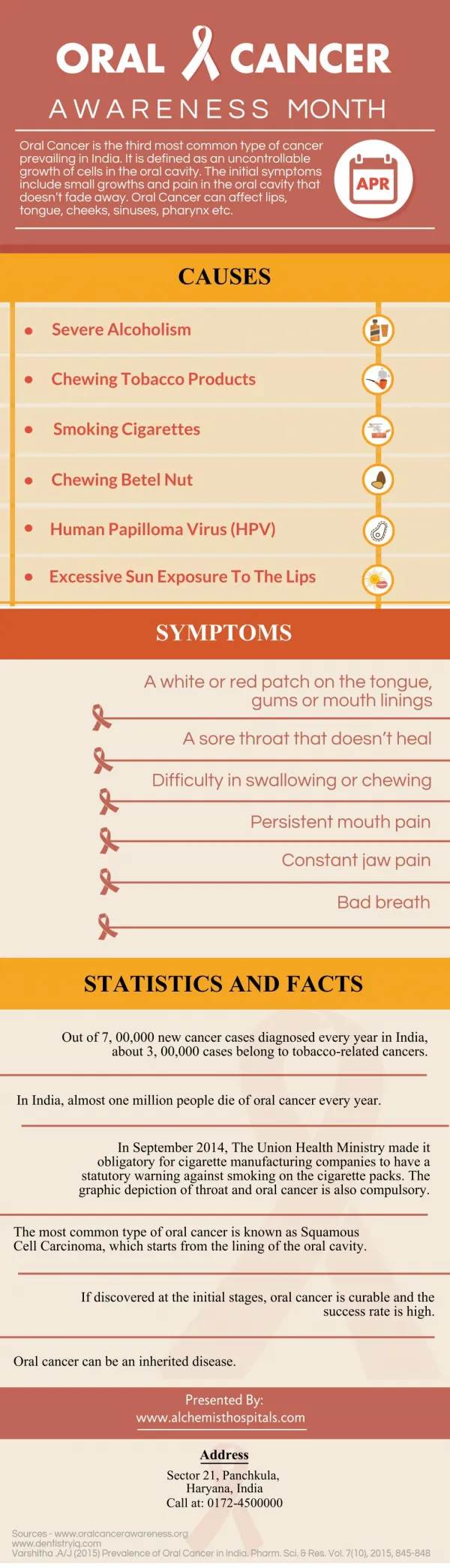
Oral Cancer-Introduction • The 6th most common malignancy within the EC although the 3rd in men (4th in women) of developing countries • The most common malignant tumour in south east asia. • 40% of all malignancies in parts of India • Commoner in males
Introduction • The prognosis for cure improves the earlier the diagnosis is made and appropriate treatment started • treatment for a small early lesion is likely to be less mutilating and have a lower morbidity than treatment for a large advanced lesion
Introduction Unlike many malignant lesions occurring elsewhere in the body oral scc can be readily observed in its early stages. There are few places in the oral cavity that a lesion can genuinely progress unnoticed by patient and clinician.
Introduction The fact that so many patients still continue to present late with advanced disease is a sad indictment of the state of medical and dental care in the UK!
Incidence and Survivalof Oral or Pharyngeal Cancer • 5410 new cases diagnosed yearly • Males 3594 • Females 1816 • 5 year survival rate: 50% UK 2007
Epidemiology Squamous cell carcinoma (scc) accounts for about 90% of all oral malignancy the remainder include salivary gland neoplasms, lymphomas and sarcomas.
Epidemiology The rate of new oral cancers would appear to be falling from its peak in 1920 to the present levels. However, there is disturbing evidence that cancers of all types including oral cancer are on the increase.
Epidemiology • there is a strong clinical impression as yet unsubstantiated that we are seeing a rise in incidence of aggressive oral scc in young patients with no accepted risk factors
Actinic Radiation Epithelial atrophy Viruses Immunosuppression Candida infection Smoking Chewing habits Alcohol Poor diet Industrial hazards Dental factors Aetiology
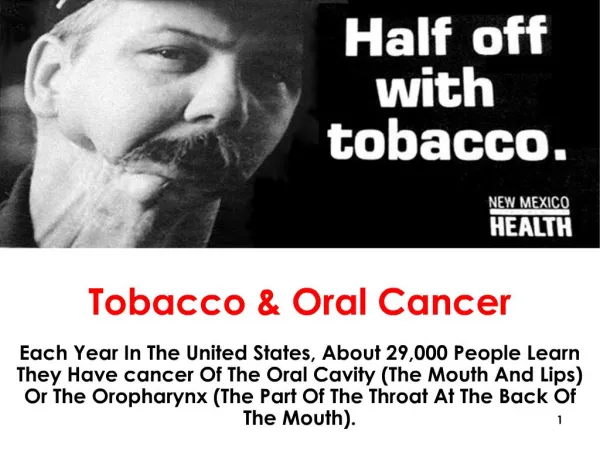
Aetiology • Smoking: Cigar and pipe smoking Vs cigarette smoking Reverse smoking • Chewing habits: Pan chewing → Leukoplakia → SCC • Alcohol: Unclear mechanism Type and quality more important than quantity
Aetiology Smoking and Alcohol synergism • Smoking alone: 9 times greater risk • Alcohol alone: 8 times greater risk • Smoking and alcohol consumption: 9+8=40!!
Aetiology • Industrial hazards: Higher incidence in textile workers • Dental factors • Actinic Radiation: SCC more common in lower lip than upper lip Lip cancer is rare in dark-skinned people • Epithelial atrophy: May enhance the absorption of carcinogens
Aetiology • Viruses: HPV particularly types 16 & 18 • Immunosuppression: Increased incidence of certain cancers in patients with renal transplants or HIV • Candidal infection: Chronic hyperplastic candidosis is premalignant
Your average oral SCC patient! • Male with carious teeth • Rarely attends dentist!!!!!! • Smokes 40-60 since ??? • Drinks cheap alcohol • Eats “junk food” on the road whilst running around in his delivery van!
Indications for urgent referral • Any unhealed ulcer for more than two weeks • Any unexplained oral bleeding • Any area of induration • Any unexplained white patch • All-red or red/white patches • Cervical nodes
Clinical presentation • Can affect any part of the oral mucosa • Sites particularly at risk vary according to aetiological factors: Europe: Tongue and lip India: Buccal mucosa
Clinical presentation • Early lesions are usually asymptomatic • May present as: a white patch a red patch an ulcer an exophytic growth
Clinical presentation • Pain may be a late feature • Advanced lesions have a very variable presentation • Bone destruction may be evident on radiographs • Teeth may become mobile • There may be altered sensation
Our Role • Patient education • Elimination of risk factors • Thorough examination • Be safe.. refer if in doubt
Examination Overview • Head and neck exam should be a routine part of dental and medical check-ups. • Take a history of alcohol and tobacco use. • Follow up on suspicious signs.
Tools and Time • Proper lighting • Dental mouth mirror • Gauze squares • Gloves • 5 minutes
Oral Lesions Suspiciousfor Oral Cancer • Homogenous leukoplakia • Leukoplakia with early squamous cell carcinoma • Nodular leukoplakia with severe epithelial dysplasia • Erythroleukoplakia with candida infection
Leukoplakia • Idiopathic white patch that cannot be wiped off the mucosa • Up to 4% risk of malignant change in 5 years • Very variable clinical presentation (homogeneous, speckled, verrucous, nodular,..etc) • Management include biopsy, conservative treatment, excision, and laser ablation
Erythroplasia (erythroplakia) • Red velvety patches • Idiopathic • Very high risk of malignant change • 70% are carcinomas in situ on first biopsy • Same management as leukoplakias
Candidal leukoplakia • Rough adherent white plaque • Typical site is buccal mucosa behind the commissures • Variable risk of malignant change • Management is with vigorous systemic antifungals
Lichen planus • Chronic inflammatory mucocutaneous disease • Unclear pathogenesis • Two distinctive clinical types • (non-erosive and erosive) • Usually bilateral distribution • Only erosive type is premalignant • Management includes biopsy and steroids
Epithelial dysplasia • Loss of tissue architecture • The degree of dysplasia is widely believed to be an important factor but there is little definitive evidence to support this assertion Early stages may be reversible !
Precancerous lesions • Risk of malignant transformation depends on: • Site • Nature of lesion • Can’t predict if the lesion will • Regress (15%) • Remain the same • Progress to cancer (4-8%)
Management of precancerous lesions • Remove the apparent cause if possible (stop smoking, antifungals) • Biopsy • Long term review
Prognosis of oral cancer STNMP system: Site Tumour size Node involvement Metastasis Pathology
Staging • T1 <2cm. T2 >2cm<4cm. T3 >4cm. T4 massive tumour with invasion • N0: No nodes • N1: ipsilateral <3cm • N2a: ipsilateral >3cm<6cm • N2b: ipsilateral multiple <6cm • N2c: Bilateral/Contralateral: <6cm • N3: any node >6cm
Investigation • Surgical biopsy, Incisional • FNA, for neck and parotid lumps • Radiographs • CT • Ultrasound esp for abdomen and liver mets.
Treatment • CURATIVE • LOCAL DISEASE CONTROL • PALIATIVE ONLY
Team Approach • Maxillofacial Surgeon • Plastic/Neuro surgery • Oncologist • Radiotherapist • Nutritionist • Speech therapist • Dentist • Maxillofacial prosthodontist
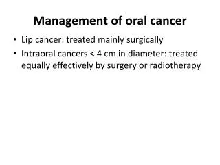
Treatment Treatment modalities: • Surgical excision • Radiotherapy • Chemotherapy?? • Surgery and radiotherapy
Surgery • Excision of the tumour with a safety margin • 1-2cm 3D margin for SCC • Intra-bony lesions require bigger margin • Partial mandibulectomy or maxillectomy with soft tissue and L.Ns
Management of the neck • Therapeutic neck dissection: When disease is obviously present in the neck and the dissection is undertaken to ablate the disease • Elective neck dissection; No obvious clinical disease in the neck but a high chance of occult disease or neck opened for access
Early Detection Saves Lives! • 5-year survival for localized disease is 76% • 5-year survival for metastatic disease is 19%
Radiation mucositis • Generalized erythematous and ulcerative response of oral mucosa • Starts the 2nd week of treatment with radiotherapy • Very painful • Secondary infection worsen the condition • subsides after the course of radiation leaving atrophic epithelium and avascular submucosa
Treatment of radiation mucositis • Sodium bicarbonate & camomile mouth-wash • Benzydamine hydrochloride MW (anti-inflammatory, anti-microbial, analgesic) • Miconazole for candidosis • Soft diet • Artificial saliva may help • PTA (polymyxin E, tobramycin, amphotericin) lozenges (reduces duration and severity)
Osteoradionecrosis • Radiation affects the vascularity of bone more susceptible to infection • Painful necrosis with sloughing of overlying soft tissues • Extraction with antibiotic cover • ORN is more likely if extraction after a long period of radiotherapy treatment
Dental practitioner role • Early detection of suspicious lesions • Prevention (e.g. stop smoking advice) • Prophylactic treatment before radiotherapy • Lifelong monitoring after radiotherapy • Prostho. treatment as part of reconstruction