Download
1 / 72
720 likes | 726 Views
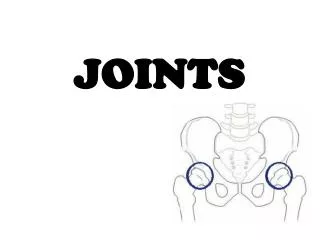
Joints. JOINT. A joint is the junction or pivot point between two or more bones. Movement of the body as a whole results from the rotation of bones about individual joints. Joints transfer and dissipate forces produced by gravity and muscle activation. ARTHROLOGY.

E N D
JOINT • A joint is the junction or pivot point between two or more bones. • Movement of the body as a whole results from the rotation of bones about individual joints. • Joints transfer and dissipate forces produced by gravity and muscle activation. Dr. Michael P. Gillespie
ARTHROLOGY • Arthrology is the study of the classification, structure, and function of joints. • Aging, long-term immobilization, trauma, and disease all affect the structure and ultimate function of joints. • These factors influence the quality and quantity of human movement. Dr. Michael P. Gillespie
Classification of Joints • Structural Classification • Presence or absence of a space (synovial cavity) • Type of Connective Tissue • Functional Classification • Relates to the degree of movement they permit. Dr. Michael P. Gillespie
CLASSIFICATION OF JOINTS BASED ON MOVEMENT POTENTIAL • Two major types of joints exist within the body: • Synarthroses • Diarthroses Dr. Michael P. Gillespie
JOINTS OF THE BODY Dr. Michael P. Gillespie
SYNARTHROSES • A synarthrosis is a junction between two bones that allows slight to essentially no movement. • Synarthroidial joints can be classified as either fibrous or cartilaginous based upon the dominant type of connective tissue. Dr. Michael P. Gillespie
TYPES OF SYNARTHROSES • Fibrous joints • Dense connective tissue (high concentration of collagen) • Sutures of the skull • Joints reinforced by an interosseous membrane (distal tibiofibular joint). • Cartilaginous joints • Flexible cartilage or hyaline cartilage • Symphysis pubis • Interbody joints of the spine • Manubriosternal joint Dr. Michael P. Gillespie
DIARTHROSES (SYNOVIAL JOINTS) • A diarthrosis is an articulation that allows moderate to extensive motion. • Possess a synovial fluid-filled cavity. • Compose the majority of the joints within the musculoskeletal system. Dr. Michael P. Gillespie
SEVEN ELEMENTS OF DIARTHRODIAL JOINTS • Articular cartilage – covers the ends and other articular surfaces of bones • Joint capsule (articular capsule) – peripheral curtain of connective tissue • Synovial membrane – dcxf vcdx d cxdfv cxfefdsex • Synovial fluid • Ligaments • Capsular ligaments • Extracapsular ligaments • Blood vessels • Sensory nerves Dr. Michael P. Gillespie
ELEMENTS OF SYNOVIAL JOINTS Dr. Michael P. Gillespie
INTRA-ARTICULAR DISCS (MENISCI) • Intra-articular discs (meninsci) – pads of fibrocartilage imposed between articular surfaces. • These pads increase congruency and improve force dispersion. • Examples of intra-articular discs • Tibiofemoral (knee) • Distal radio-ulnar • Sternoclavicular • Acromioclavicular • Temporomandibular • Apophyseal (variable) Dr. Michael P. Gillespie
PERIPHERAL LABRUM • A peripheral labrum of fibrocartilage extends from the body rims of the glenoid fossa to the shoulder and the acetabulum of the hip. • These structures deepen the concave surface of the joint. • These structures support and thicken the attachment of the joint capsule. Dr. Michael P. Gillespie
FAT PADS • Fat pads thicken the joint capsule, causing the inner surface of the capsule to fill nonarticulating joint spaces formed by incongruent bony contours. • They are prominent in the elbow and knee joints. • Enlarged and inflamed fat pads can adversely affect the biomechanics of the joint. Dr. Michael P. Gillespie
BURSAE • A bursa is an extension or outpouching of the synovial membrane of a diarthroidial joint. • Bursae are filled with synovial fluid and exists in areas of potential stress. • Bursae help to absorb force and protect periarticular connective tissues, including bone. Dr. Michael P. Gillespie
SYNOVIAL PLICA • Synovial plicae (synovial folds, synovial redundancies, or synovial fringes) are slack, overlapped pleats of tissue composed of the innermost layers of the joint capsule. • They are found in joints with large capsular surface area such as the knee and elbow. • They increase the synovial surface area and allow full joint motion without undue tension on the synovial lining. • Folds that are thickened or adhered due to inflammation can alter the joint biomechanics. Dr. Michael P. Gillespie
Structural Classification of Joints • Fibrous Joints • Fibrous CT • Lack a synovial cavity • Cartilaginous Joints • Cartilage • Lack a synovial cavity • Synovial Joints • Have a synovial cavity • Dense irregular CT • Often associated with accessory ligaments Dr. Michael P. Gillespie
Functional Classification of Joints • Synarthrosis (syn = together) • Immovable joint • Amphiarthrosis (amphi = on both sides) • A slightly moveable joint • Diarthrosis (moveable joint) • A freely moveable joint • Synovial joints Dr. Michael P. Gillespie
Fibrous Joints • Lacks a synovial cavity • Little or no movement Dr. Michael P. Gillespie
Fibrous Joints • Sutures • Immovable • Synostosis – suture that is replaced by bone in the adult • Syndesmoses • Slightly moveable (amphiarthrosis) • Ligament • Interosseous membrane • Gomphoses • Dentoalveolar joint Dr. Michael P. Gillespie
Cartilaginous Joints • Lacks a synovial cavity • Allows little or no movement • Synchondroses • Epiphyseal plate • Symphyses • Pubic symphisis • Intervertebral discs Dr. Michael P. Gillespie
Synovial Joints • Synovial (Joint) Cavity – space btwn. Bones • Freely moveable • The bones are covered by hyaline cartilage • Contains the following: • Articular capsule • Synovial fluid • Accsessory ligaments and articular discs Dr. Michael P. Gillespie
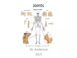
Classification of Synovial Joints Based on Mechanical Analogy • Hinge joint • Pivot joint • Ellipsoid joint • Ball-and-socket joint • Plane joint • Saddle joint • Condyloid joint Dr. Michael P. Gillespie
HINGE JOINT • Primary Angular Motions • Flexion and extension only • Mechanical Analogy • Door hinge • Anatomic Examples • Humero-ulnar joint • Interphalangeal joint Dr. Michael P. Gillespie
HINGE JOINT Dr. Michael P. Gillespie
PIVOT JOINT • Primary Angular Motions • Spinning of one member around a single axis of rotation • Mechanical Analogy • Doorknob • Anatomic Examples • Humeroradial joint • Atlanto-axial joint Dr. Michael P. Gillespie
PIVOT JOINT Dr. Michael P. Gillespie
ELLIPSOID JOINT • Primary Angular Motions • Biplanar motion (flexion-extension and abduction-adduction) • Mechanical Analogy • Flattened convex ellipsoid paired with a concave trough • Anatomic Examples • Radiocarpal joint Dr. Michael P. Gillespie
ELLIPSOID JOINT Dr. Michael P. Gillespie
BALL-AND-SOCKET JOINT • Primary Angular Motions • Triplanar motion (flexion-extension, abduction-adduction, and internal-external rotation) • Mechanical Analogy • Spheric convex surface paired with a concave cup • Anatomic Examples • Glenohumeral joint • Coxofemoral (hip) joint Dr. Michael P. Gillespie
BALL-AND-SOCKET JOINT Dr. Michael P. Gillespie
PLANE JOINT • Primary Angular Motions • Slide (translation) or combined slide and rotation • Mechanical Analogy • Relatively flat surfaces apposing each other, like a book on a table • Anatomic Examples • Carpometacarpal joints (digits II to IV) • Intercarpal joints • Intertarsal joints Dr. Michael P. Gillespie
PLANE JOINT Dr. Michael P. Gillespie
SADDLE JOINT • Primary Angular Motions • Biplanar motion • Spin between bones is possible, but may be limited by interlocking nature of joint • Mechanical Analogy • Each member has a reciprocally curved concave and convex surface oriented at right angles to the other, like a horse rider and a saddle • Anatomic Examples • Carpometarcarpal joint of the thumb • Sternoclavicular joint Dr. Michael P. Gillespie
SADDLE JOINT Dr. Michael P. Gillespie
CONDYLOID JOINT • Primary Angular Motions • Biplanar motion • Either flexion-extension and abduction-adduction, or flexion-extension and axial rotation (internal-external rotation) • Mechanical Analogy • Mostly spheric convex surface that is enlarged in one dimension like a knuckle; paired with a shallow concave cup • Anatomic Examples • Metacarpophalangeal joint • Tibiofemoral (knee) joint Dr. Michael P. Gillespie
CONDYLOID JOINT Dr. Michael P. Gillespie
SIMPLIFYING THE CLASSIFICATION OF SYNOVIAL JOINTS • Two articular forms based upon true movement of the joint. • Ovoid joint • Saddle joint • Essentially all synovial joints of the body with the notable exception of planar joints can be categorized under this scheme. Dr. Michael P. Gillespie
OVOID JOINT • An ovoid joint has paired mating surfaces that are imperfectly spheric, or egg-shaped, with adjacent parts possessing a changing surface curvature. • The articular surface of one bone is convex and of the other is concave. • Most joints of the body are of this variety. Dr. Michael P. Gillespie
SADDLE JOINT • A saddle joint consists of paired convex and concave surfaces oriented at approximately 90 degrees to each other. • Each member has a reciprocally curved concave and convex surface oriented at right angles to the other, like a horse rider and a saddle. Dr. Michael P. Gillespie
BASIC SHAPES OF JOINT SURFACES Dr. Michael P. Gillespie
BIOLOGICAL MATERIALS OF PERIARTICULAR CONNECTIVE TISSUES • Fibrous Proteins • Collagen (type I and II) • Ground Substance • Glycosaminoglycans • Water • Solutes • Cells • Fibroblasts • Chondrocytes Dr. Michael P. Gillespie
TYPES OF COLLAGEN IN PERIARTICULAR CONNECTIVE TISSUES • Type I • Thick, rugged fibers that elongate when stretched • Present in ligaments, tendons, fascia, and fibrous joint capsules • Type II • Thinner fibers than type I • Provide a framework for maintaining the general shape and consistency of structures, such as hyaline cartilage Dr. Michael P. Gillespie
Types of Movements at Synovial Joints • Gliding • Simple back and forth movement, limited in range, planar joints • Angular Movements • Increase or decrease in the angle btwn. bones • Rotation • Bone revolves around a longitudinal axis • Special Movements Dr. Michael P. Gillespie
Angular Movements • Flexion, extension, lateral flexion, hyperextension • Abduction, adduction, and circumduction Dr. Michael P. Gillespie
Rotation • Medial (internal) rotation • Lateral (external) rotation Dr. Michael P. Gillespie
Special Movements • Elevation • Depression • Protraction • Retraction • Inversion Dr. Michael P. Gillespie
Special Movements • Eversion • Dorsiflexion • Plantar flexion • Supination • Pronation • Opposition Dr. Michael P. Gillespie
Dislocation • Luxation – displacement of a bone from a joint • Causes tearing or ligaments, tendons, and articular capsules • Subluxation • Incomplete dislocation Dr. Michael P. Gillespie