Download
1 / 41
410 likes | 547 Views
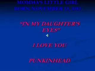
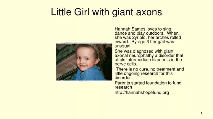
Hannah Sames loves to sing, dance and play outdoors. When she was 2yr old, her arches rolled inward. By age 3 her gait was unusual. She was diagnosed with giant axonal neurophathy a disorder that affcts intermediate filaments in the nerve cells.

E N D
Hannah Sames loves to sing, dance and play outdoors. When she was 2yr old, her arches rolled inward. By age 3 her gait was unusual. She was diagnosed with giant axonal neurophathy a disorder that affcts intermediate filaments in the nerve cells. There is no cure, no treatment and little ongoing research for this disorder Parents started foundation to fund research http://hannahshopefund.org Little Girl with giant axons
Cell Division and Death Normal growth and development require an intricate interplay between the rates of two processes Mitosis – Cell division - Produces two somatic cells from one Apoptosis – Cell death - Precise genetically-programmed sequence that carves toes and fingers during embryonic development Figure 2.3
Figure 2.13 Figure 2.12
The Cell Cycle The sequence of events associated with cell division G phase: Gap for growth S phase: DNA synthesis M phase: Mitosis (nuclear division) Cytokinesis: Cell division Figure 2.14 Figure 2.3
Stages of the Cell Cycle Interphase - Prepares for cell division - Replicates DNA and subcellular structures - Composed of G1, S, and G2 - Cells may exit the cell cycle at G1 or enter G0, a quiescent phase Mitosis – Division of the nucleus Cytokinesis – Division of the cytoplasm Figure 2.3
Replication of Chromosomes Chromosomes are replicated during S phase prior to mitosis The result is two sister chromatids held together at the centromere Figure 2.15 Figure 2.3
Mitosis Used for growth, repair, and replacement Consists of a single division that produces two identical daughter cells A continuous process divided into 4 phases - Prophase - Metaphase - Anaphase - Telophase Figure 2.3
Mitosis in a Human Cell Figure 2.15 Figure 2.16
Prophase Replicated chromosomes condense Microtubules organize into a spindle Nuclear envelope and nucleolus break down Figure 2.3 Figure 2.16
Metaphase Chromosomes line up on the cell’s equator Spindle microtubules are attached to centromeres of chromosomes Figure 2.3 Figure 2.16
Anaphase Centromeres divide Chromatids separate and become independent chromosomes - They move to opposite ends of the cell Figure 2.3 Figure 2.16
Telophase Chromosomes uncoil Spindle disassembles Nuclear envelope reforms Figure 2.3 Figure 2.16
Cytokinesis Cytoplasmic division occurs after nuclear division is complete Organelles and macromolecules are distributed between the two daughter cells Microfilament band contracts, separating the two cells Figure 2.3
Mitosis Animation Please note that due to differing operating systems, some animations will not appear until the presentation is viewed in Presentation Mode (Slide Show view). You may see blank slides in the “Normal” or “Slide Sorter” views. All animations will appear after viewing in Presentation Mode and playing each animation. Most animations will require the latest version of the Flash Player, which is available at http://get.adobe.com/flashplayer. Figure 2.3
Cell Cycle Control Checkpoints ensure that mitotic events occur in the correct sequence Internal and external factors are involved Many types of cancer result from faulty checkpoints Figure 2.3
Cell Cycle Control Figure 2.17 Figure 2.16
Telomeres Located at the ends of the chromosomes Contain hundreds to thousands of repeats of a 6-base DNA sequence Most cells lose 50-200 endmost bases after each cell division After about 50 divisions, shortened telomeres signal the cell to stop dividing Sperm, eggs, bone marrow, and cancer cells produce telomerase that prevent shortening of telomeres Figure 2.3
Apoptosis Begins when a cell receives a “death signal” Killer enzymes called caspases are activated -Destroy cellular components Phagocytes digest the remains Dying cell forms bulges called blebs Figure 2.3
Programmed cell death is part of normal development Figure 2.19 Mitosis and apotosis work together to form functional body Cancer can result from too much mitosis, too little apotosis Figure 2.18
Cell-to-Cell Interactions Make multicellular life possible Two broad types 1) Signal transduction 2) Cellular adhesion Defects cause certain inherited disorders Figure 2.3
Signal Transduction The process of transmitting a signal from the environment to a cell - Receptor binds to “first messenger” - Interacts with regulator - Causes enzyme to produce “second messenger” - Elicits cellular response, which is typically enzyme activation - Amplification due to cascade Figure 2.3
Defects in signal transduction underlie many inherited disorders. For example neurofibromatosis type 1 in which benign tumors grow in the nervous tissue especially under the skin Signal Transduction
Signal Transduction Figure 2.20 Figure 2.19
Second Messenger Animation Please note that due to differing operating systems, some animations will not appear until the presentation is viewed in Presentation Mode (Slide Show view). You may see blank slides in the “Normal” or “Slide Sorter” views. All animations will appear after viewing in Presentation Mode and playing each animation. Most animations will require the latest version of the Flash Player, which is available at http://get.adobe.com/flashplayer. Figure 2.3
Cellular Adhesion A precise sequence of interactions among proteins that connect cells Example = Inflammation - Three types of cellular adhesion molecules (CAMs) help guide WBCs to the injured area - Secretins, integrins, and adhesion receptor proteins Figure 2.3
Cellular Adhesion Figure 2.21 Figure 2.20
Stem Cells A stem cell divides by mitosis - Produces daughter cells that retain the ability to divide and some that specialize Progenitor cells do not have the capacity of self-renewal Figure 2.3 Figure 2.22
Stem Cells All cells in the human body descend from stem cells via mitosis and differentiation Cells differentiate down cell lineages by differential gene expression Stem cells are present throughout life and provide growth and repair Figure 2.3
Figure 2.23 Figure 2.3
Stem Cells Stem cells and progenitor cells are described in terms of their developmental potential Totipotent – Can give rise to every cell type Pluripotent – Have fewer possible fates Multipotent – Have only a few fates Figure 2.3
Stem Cells in Health Care There are 3 general sources of human stem cells 1) Embryonic stem cells – Created in a lab dish using the inner cell mass (ICM) of an embryo 2) Induced pluripotent stem (iPS) cells – Somatic cells reprogrammed to differentiate into any of several cell types 3) Adult stem cells – Tissue-specific or somatic stem cells Figure 2.3
Stem Cells in Health Care Figure 2.24 Figure 2.24
Stem Cell Applications Stem cells are being used in four basic ways 1) Discovery and development of drugs 2) Observing the earliest sign of disease 3) Treatment of disease via implants and transplants 4) Stimulating stem cells in the body via the introduction of reprogramming proteins Figure 2.3
Reproductive cloning: also called somatic cell nuclear transfer • oocyte is retrieved • Enucleation: the removal of nucleus from the cell • somatic cell nuclear transfer from donor to cell ( in Dolly this was a mammillary gland cell from a 6 yr old sheep • activation of embryo to develop • embryo transfer to surrogate mom
Therapeutic cloning: • #1-4 same • Then the clone of the donors embryo grows in a Petri dish for 6 days ( cells in the blastocyst stage) ( window for use is 10 days…on day 6 this means cells are viable for 4 more days) ( on day 11 they change and are no longer in blastocyst stage) • The embryonic stem cells are then removed from the embryo and used to produce cells for therapy, the embryo is destroyed in this process. • These cells can be used because they are pluripotent stem cells meaning they can be programmed to become any of the 220 different types of cells found in the human body
Alternative to embryo destruction • The formation of ethical pluripotent stem cells: (those that arise without the destruction of an embryo)such as IPs …this can be done by: • biopsy of 8 cell embryo ( remove one cell) • ANT: altered nuclear transfer • living cells from dead embryo • parthenogenesis • reprogramming cells with a new signal ( cells such as skin cells are used and go through reproductive steps #2- 5 with 5 being the donors needed organs) • Ips: means induced pluripotent stem cells
Stem Cell Applications Figure 2.25 Figure 2.3
Cloning from differentiated somatic cells. Ian Wilmut and his team of researchers at Roslin Institute in Scotland cloned Dolly, the first mammal to be cloned from a somatic cell.
Nuclear transfer method of producing clones Animal Cloning
Four reasons why genetically identical animals are not phenotypically identical. Mitochondria Immune response Epigenetics Embryo development
Cloning at Texas A&M • World leader in producing the most number of clones from different species • Cattle • Second Chance (1999) • 862 (2000) • Goat – Second Addition (2001) • Pig litter (2001) • Cat - Cc (2001) • Deer – Dewey (2003) • Horse – Paris Texas (2005)